Method for detecting leptospira by multiple cross amplification in combination with nano-biosensor

A biosensor and leptospira technology, applied in the field of microbial detection, can solve the problems of complex operation steps and dependence on reagents, and achieve the effect of fast detection speed and excellent detection sensitivity

Active Publication Date: 2019-01-01
贵州省疾病预防控制中心
View PDF2 Cites 4 Cited by
  • Summary
  • Abstract
  • Description
  • Claims
  • Application Information

AI Technical Summary

Problems solved by technology

However, these constant temperature technologies require multiple enzymes to work simultaneously to achieve nucleic acid amplification, relying on expensive reagents and complicated

Method used


Image

  • Method for detecting leptospira by multiple cross amplification in combination with nano-biosensor
  • Method for detecting leptospira by multiple cross amplification in combination with nano-biosensor
  • Method for detecting leptospira by multiple cross amplification in combination with nano-biosensor

Examples


Embodiment 1

[0048] The feasibility of embodiment 1.MCDA-LFB amplification

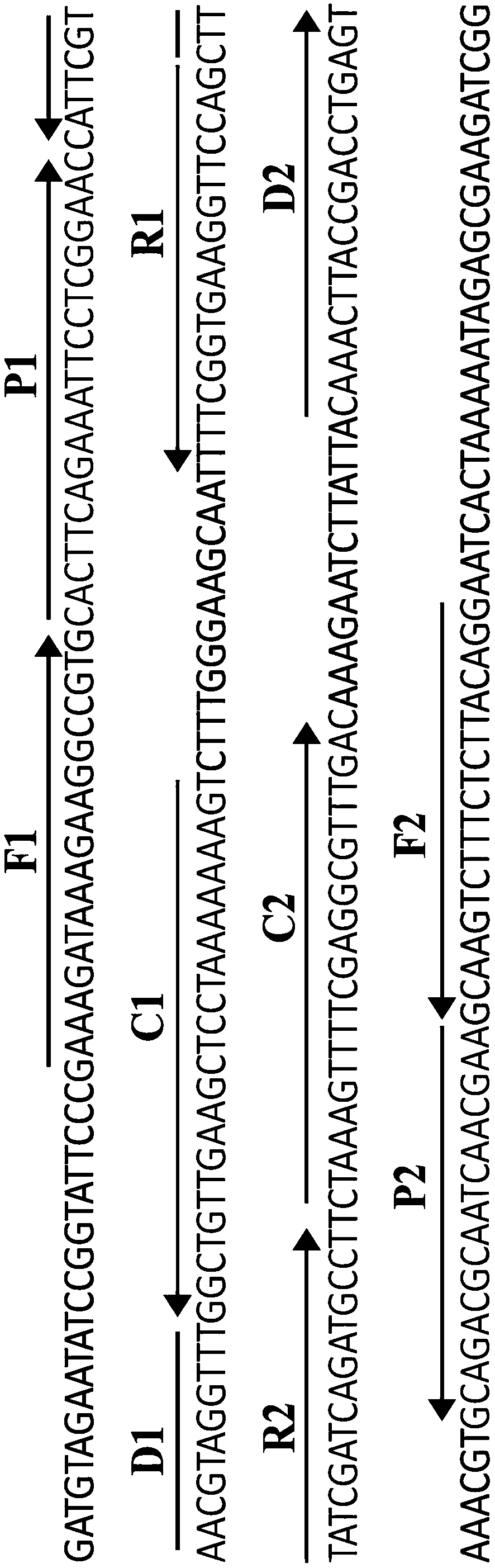
[0049] Standard MCDA reaction system: the concentration of cross primers CP1 and CP2 is 60 pmol, the concentration of displacement primers F1 and F2 is 10 pmol, the concentration of amplification primers R1, R2, D1* and D2 is 30 pmol, and the concentration of amplification primers C1* and C2 The concentration is 20pmol, 10mM Betain, 6mM MgSO 4 , 1 mM dNTP, 12.5 μL of 10×Bst DNA polymerase buffer, 10 U of strand-displacing DNA polymerase, 1 μL of template, and add deionized water to 25 μl. The entire reaction was thermostated at 64°C for 35 minutes.

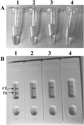
[0050] After MCDA amplification, two detection methods are used to discriminate MCDA amplification. First, a visible dye (such as MG reagent, leuco malachite green visual reagent) is added to the reaction mixture, and the color of the positive reaction tube changes from colorless to It is bright green, and the negative reaction tube remains colorless. Secondly, MCD...

Embodiment 2

[0053] Embodiment 2. measure the optimal reaction temperature of MCDA technology

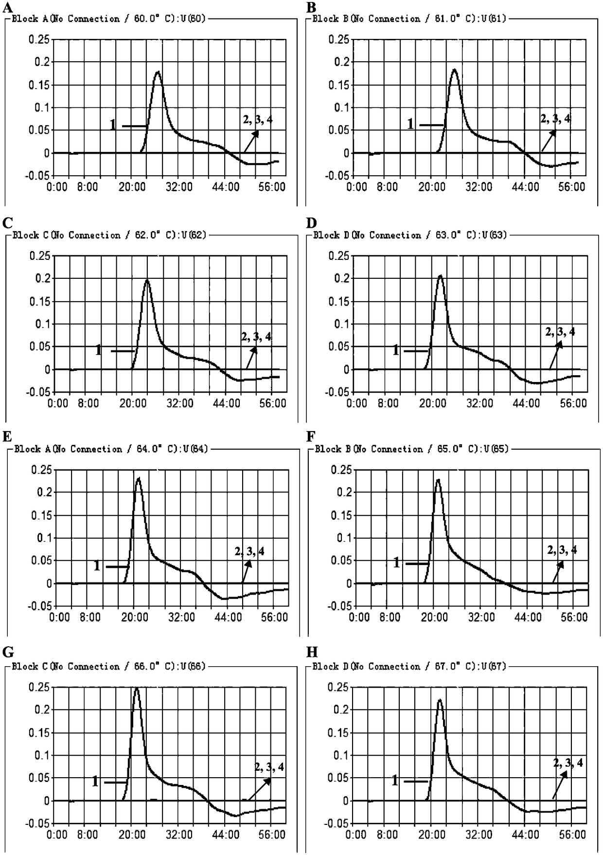
[0054] Under standard reaction system conditions, the DNA template for pathogenic Leptospira and the corresponding MCDA primer designed were added, and the template concentration was 10 pg / ul. The reaction was carried out under constant temperature conditions (60-67°C), and the results were detected by a real-time turbidimeter, and different dynamic curves were obtained at different temperatures, see image 3 . 61-64°C ( image 3 Middle B-E) are recommended as the optimal reaction temperature for MCDA primers. In the follow-up verification of the present invention, 62° C. was selected as the constant temperature condition for MCDA amplification. image 3 It represents the temperature dynamic curve of MCDA primers designed for the lipL41 gene sequence to detect pathogenic Leptospira.

Embodiment 3

[0055] Embodiment 3.MCDA-LFB detects the sensitivity of single target

[0056] After using the serially diluted pathogenic Leptospira genomic DNA to carry out the standard MCDA amplification reaction, the LFB detection showed that the detection range of MCDA-LFB was 10 ng-10 fg, and LFB appeared red lines in the TL and CL regions ( Figure 4 Middle A). When the amount of genomic template in the reaction system is reduced to below 10fg, LFB only appears a red line in the CL area, indicating a negative result ( Figure 4 LFB6-8 in A). Figure 4 Middle A is the use of LFB to visually read the MCDA amplification results; Figure 4 LFB 1 to 8 in A respectively represent that the template amount of pathogenic Leptospira is 1x10 5 genomic equivalents(GEs) / reaction; 1x10 4 GEs / reaction; 1x10 3 GEs / reaction; 1x10² GEs / reaction; 1x10 1 GEs / reaction; 1x10 0 GEs / reaction; 1x10 -1 GEs / reaction; 1 µl double distilled water.

[0057] In order to further verify the sensitivity of MCD...


PUM


Abstract

The invention discloses a method for detecting leptospira by multiple cross amplification in combination with a polymer nano-biosensor. The method uses specially designed multi-cross displacement amplification primers, and also provides a primer formed by labeling the 5' end of the amplification primer C1 with biotin and a primer formed by labeling the 5' end of the amplification primer D1 with hapten. A product of the amplification of the LipL41 gene of pathogenic leptospira in the method can be visually detected by the polymer nano-biosensor. The method is convenient, fast, sensitive and specific, and is suitable for the specific detection of pathogenic leptospira.

Description

technical field [0001] The invention discloses a method for detecting pathogenic Leptospira, belonging to the field of microorganism detection. Background technique [0002] Leptospira is a kind of slender bacteria with different lengths, generally 6-20um, 0.1-0.2um wide, with fine and regular spiral bacteria, one or both ends of the bacteria are bent into a hook shape, often "c", "s" and other shapes. Under the dark field microscope, the leptospira can be seen as a string of shiny microbeads, which can flex, move back and forth or rotate rapidly around the long axis. Under the electron microscope, the leptosome is a cylindrical structure, the outermost layer is the sheath, which is composed of lipopolysaccharide and protein, the inner part is the cell wall, and the inner part is the serous membrane. The central axis of the formation is located on the side of the thallus. Leptospira is formed by winding the central axis of the entire cylindrical bacterium, and the cell wa...

Claims


Application Information

Patent Timeline
IPC IPC(8): C12Q1/6844C12Q1/04C12N15/11
CPCC12Q1/6844C12Q2565/607C12Q2537/1376C12Q2531/119Y02A50/30
Inventor 李世军刘英
Owner 贵州省疾病预防控制中心
Why Eureka
Try Eureka