Method for rapidly and quantitatively detecting lactobacillus rhamnosus phages

A technology of Lactobacillus rhamnosus and quantitative detection method, which is applied in the field of microorganisms, can solve the problems of no corresponding method for phage detection, high detection cost, and long detection cycle, so as to quickly determine the source and route of phage contamination, and is easy to popularize and use , Improve the effect of timeliness

Active Publication Date: 2019-01-18
YANGZHOU UNIV
View PDF2 Cites 5 Cited by
  • Summary
  • Abstract
  • Description
  • Claims
  • Application Information

AI Technical Summary

Problems solved by technology

The conventional double-layer agar plate method is currently the most widely used phage detection method, but the detection cycle is long, usually requiring more than 24 hours of cultivation, and it is difficult to meet the requirements of fermentation production in terms of timeliness
In recent years, PCR technology has been applied to the rapid detection of Lactobacillus phages and nisin streptococcal phages. Althoug

Method used


Image

  • Method for rapidly and quantitatively detecting lactobacillus rhamnosus phages
  • Method for rapidly and quantitatively detecting lactobacillus rhamnosus phages
  • Method for rapidly and quantitatively detecting lactobacillus rhamnosus phages

Examples


Embodiment 1

[0030] Example 1 Preparation of immunomagnetic beads and capture of live cells of Lactobacillus rhamnosus

[0031] The preparation steps of immunomagnetic beads are as follows: 2 mg (200 μL) carboxy-modified magnetic nanobeads (MNB) were placed in a 1.5 mL centrifuge tube, washed twice with 500 μL MEST buffer, and then suspended in 500 μL EDC-NHSS solution. After activation at 37°C for 30 min, MNB was collected by magnetic separation, suspended with 500 μL PBST, washed 3 times, and added to a new sterile 1.5 mL centrifuge tube. Add the specific polyclonal antibody (PcAb-SpaA) (PcAb-SpaA) (100 μL) of Lactobacillus rhamnosus pilus subunit to the solution, incubate at 37°C for 3 h, and discard the supernatant. The PcAb-SpaA-coupled MNB (PcAb-SpaA-MNB) was blocked in PBST containing 1% BSA at 37°C for 45 min, washed 3 times with PBST, and washed with 0.02% NaN 3 Resuspend with 0.5% BSA in PBST (200 μL) to a final concentration of 10 mg / ml, and store at 4°C for later use.

[0032...

Embodiment 2

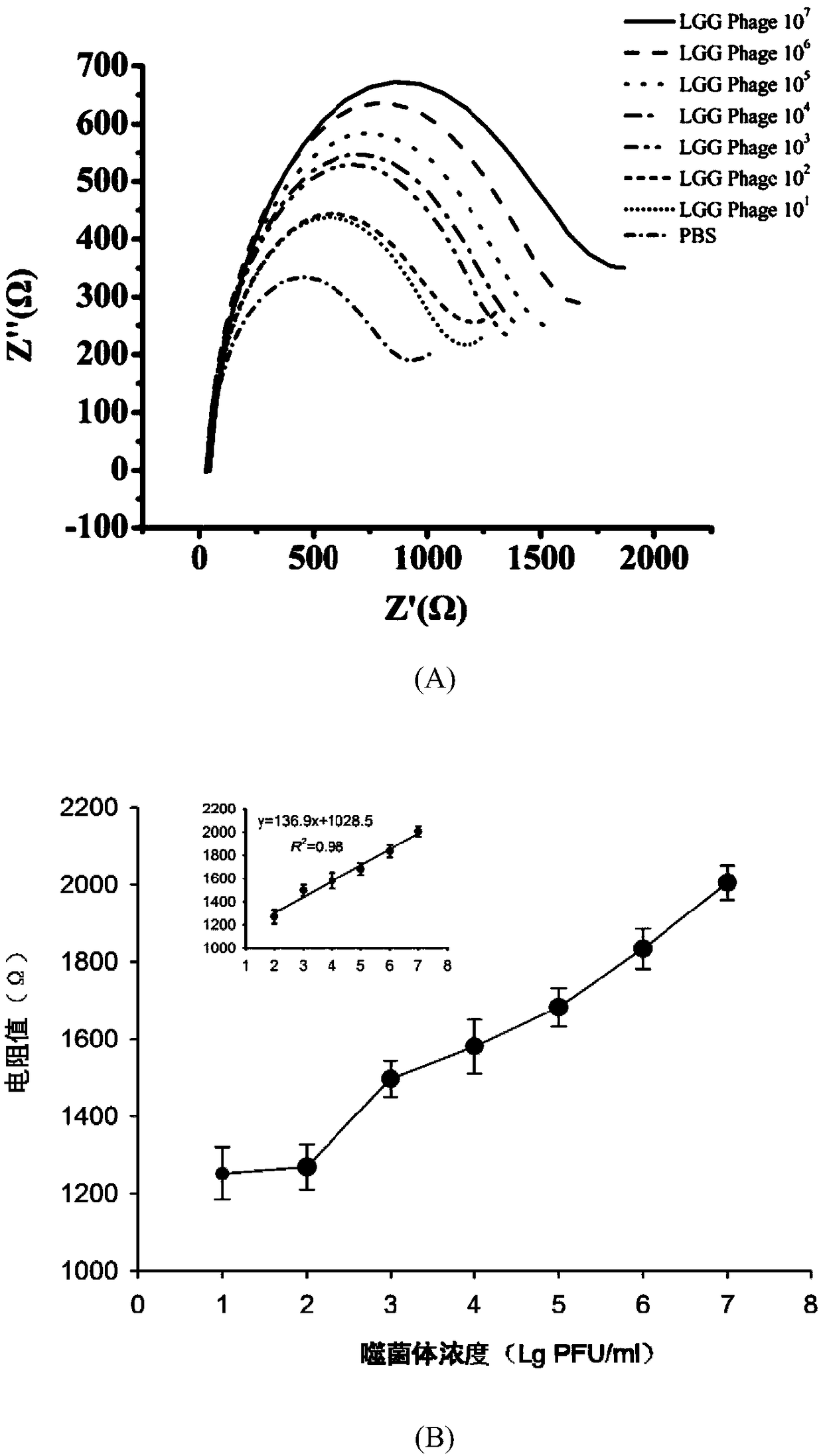
[0035] Example 2 Phage Capture and Electrochemical Signal Detection

[0036] Phage capture Take 100 μL of 109 PFU / mL Lactobacillus rhamnosus (LGG strain) phage diluted to 10 with PBS 7 PFU / mL, 10 6 PFU / mL, 10 5 PFU / mL, 10 4 PFU / mL, 10 3 PFU / mL, 10 2 PFU / mL to prepare phage gradient samples, and do 3 parallels for each concentration; 9 PFU / mL Enterobacter sakazakii phage, diluted to 10 with PBS 7 PFU / mL, 10 6 PFU / mL, 10 5 PFU / mL, 10 4 PFU / mL, 10 3 PFU / mL, 10 2 PFU / mL, three parallels were done for each concentration; PBS without phage was used as a blank control. Take 100 μL of live cell suspension of Lactobacillus rhamnosus and add it to 900 μL of the above-mentioned phage gradient dilution sample, mix well, and let stand at room temperature for 15 minutes for adsorption; take 10 μL of immunomagnetic beads prepared according to (1) and add PcAb-SpaA-MNB Add to each sample, mix well, incubate at 37°C for 60 min; discard the supernatant after magnetic fi...

Embodiment 3

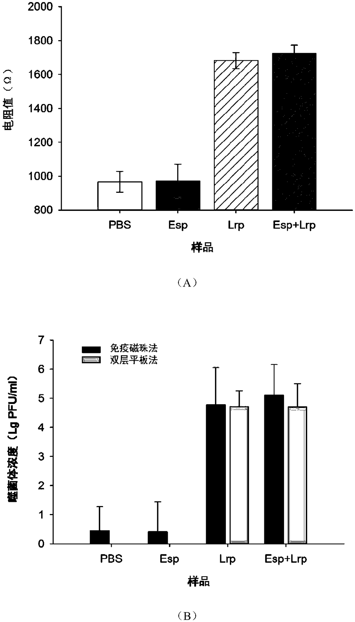
[0039] Embodiment 3 simulated sample detection result

[0040] Take 10μL concentration of 10 7 PFU / mL of Lactobacillus rhamnosus phage and 10 μL of 10 7 Mix PFU / mL Enterobacter sakazakii phage, add 1ml MRS medium and mix thoroughly to make a sample, use MRS without Lactobacillus rhamnosus phage as a negative control; use the traditional double-layer plate method to determine the phage content, specific steps: Take 10 μL of the sample with sterile SM buffer for serial dilution, take 100 μL of the appropriate dilution and mix it with 100 μL of Lactobacillus rhamnosus cultivated overnight at room temperature for 15 min, add 5 mL of MCM semi-solid medium, Quickly pour it onto the bottom plate, wait for the medium to fully solidify, incubate at a constant temperature of 37°C until clear phage plaques appear, and count the number of phage plaques.

[0041] Take 100 μL of live cell suspension of Lactobacillus rhamnosus and add 900 μL of the above sample, mix well, let stand at ro...


PUM


Abstract

The invention relates to the technical field of microorganisms, in particular to a method for rapidly and quantitatively detecting lactobacillus rhamnosus phages. According to the method, a lactobacillus rhamnosus pili subunit SpaA specific polyclonal antibody is coupled to nano-magnetic beads to form specific immunomagnetic beads; phage particles in a sample are adsorbed by using lactobacillus rhamnosus viable cells, and then are re-suspended in a buffer solution after being centrifuged and washed; and after the immunomagnetic beads are used to capture a lactobacillus rhamnosus-phage compound, and magnetic field separation and PBS washing are performed, a magnetic glassy carbon electrode is taken as a working electrode, a lead wire and a saturated calomel electrode are taken as a counterelectrode and a reference electrode, the difference between resistance values in the sample and a contrast is determined, and the phages in the sample can be rapidly and quantitatively detected. The method is simple and convenient in operation, free of culture, short in time and convenient to popularize and use, is suitable for detection of the phages in an environment and raw materials in high-density fermentation preparation of lactobacillus rhamnosus, and is convenient for rapidly determining pollution source and way; and comprehensive prevention and control measures are adopted.

Description

technical field [0001] The invention relates to the technical field of microorganisms, in particular to a rapid quantitative detection method for Lactobacillus rhamnosus phage. Background technique [0002] Lactobacillus rhamnosus ( Lactobacillus rhamnosus ) is currently recognized as a probiotic lactobacillus, which has physiological functions such as maintaining the balance of intestinal microecology, inhibiting the growth of harmful bacteria, and eliminating allergies. It has been widely used in food supplements and probiotic products. The preparation of high-density Lactobacillus rhamnosus powder by fermentation is the basis of functional dairy products and probiotic health care products. At present, this industry has produced huge economic and social benefits. However, during the fermentation process, lactic acid bacteria are often infected by phages, resulting in weakened activity, slow fermentation, and reduced product quality, resulting in economic losses. Therefor...

Claims


Application Information

Patent Timeline
IPC IPC(8): G01N33/569G01N33/543
CPCG01N33/54326G01N33/54346G01N33/5695
Inventor 杨振泉时秒高璐胡钦饶胜其郑香峰
Owner YANGZHOU UNIV
Why Eureka
Try Eureka