Method for separating and preparing exosome

A technology of exosomes and liquids, applied in the biological field, can solve problems such as pollution, multiple impurities, residues, etc.

Inactive Publication Date: 2019-01-25
INST OF HEMATOLOGY & BLOOD DISEASES HOSPITAL CHINESE ACADEMY OF MEDICAL SCI & PEKING UNION MEDICAL COLLEGE
View PDF3 Cites 4 Cited by
  • Summary
  • Abstract
  • Description
  • Claims
  • Application Information

AI Technical Summary

Problems solved by technology

However, the impurity removal process is relatively simple, and there will be more impurities remaining, which will cause pollution to subsequent experiments, such as residual cellular proteins, nucleic acids and other components

Method used


Image

  • Method for separating and preparing exosome
  • Method for separating and preparing exosome
  • Method for separating and preparing exosome

Examples


Embodiment 1

[0050] Example 1: A method for the isolation and preparation of exosomes

[0051] 1. Materials

[0052] Peripheral blood: from discarded samples after clinical examination of patients

[0053] Bone marrow plasma: from discarded specimens after clinical examination of patients

[0054] Reagent XBP, Reagent XWP, membrane adsorption column exoEasy Midi Spin Columns, column Spin Column comes from the kit QIAGEN exoRNeasy Serum / PlasmaMidi Kits catNo: 77044.

[0055] Reagent XE: QIAGEN Lot No.160011905

[0056] Exosome RNA extraction kit: exoRNeasy Serum / Plasma Midi Kit, Qiagen, Germany, cat.No.77044

[0057] 2. Method

[0058] 1. Centrifuge to remove cellular impurities: Centrifuge peripheral blood or bone marrow plasma at 4°C and 2500g for 10 minutes to remove the influence of cellular impurities such as red blood cells and white blood cells, discard the precipitate and retain the supernatant, and transfer 500 μl of the supernatant to a new 1.5 ml EP tube.

[0059] 2. C...

Embodiment 2

[0069] Example 2: Extraction of exosome total RNA

[0070] 1. Materials

[0071] Example 1 Obtaining a liquid sample containing exosomes

[0072] Exosome RNA extraction kit: exoRNeasy Serum / Plasma Midi Kit, Qiagen, Germany, cat.No.77044

[0073] 2. Method

[0074] 1. Add 700 μl QIAzol to the exosome-containing resuspension obtained above

[0075] 2. Vortex the EP tube for 5 seconds, let stand at room temperature (15°C-25°C) for 5 minutes, add 90 μl chloroform and shake vigorously up and down for 15 seconds, and let stand at room temperature for 2-3 minutes;

[0076] 3. Centrifuge at 12000g for 15min at 4°C;

[0077] 4. Transfer the upper aqueous phase to a new EP tube to avoid sucking into the middle organic layer;

[0078] 5. Add twice the volume of 100% ethanol to the upper layer of water (for example, add 800 μl of 100% ethanol to the upper layer of 400 μl of water), mix well by blowing up and down, draw 700 μl of liquid and add it to the column (RNeasy MinElute Spin C...

Embodiment 3

[0087] Example 3. Detection of RNA quality in exosomes and exosomes

[0088] 1. Materials

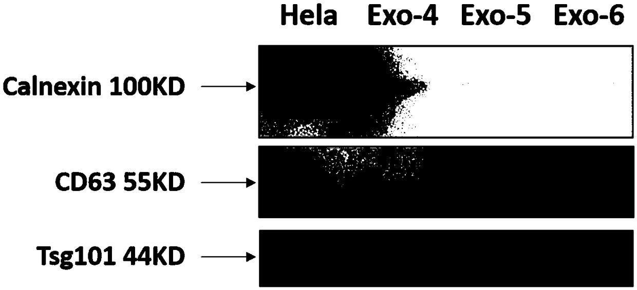
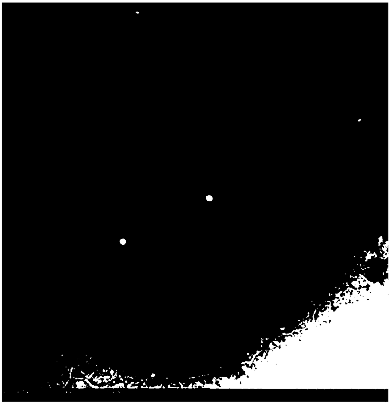
[0089] The exosome sample obtained in Example 1 (the sample number is Exo-6)

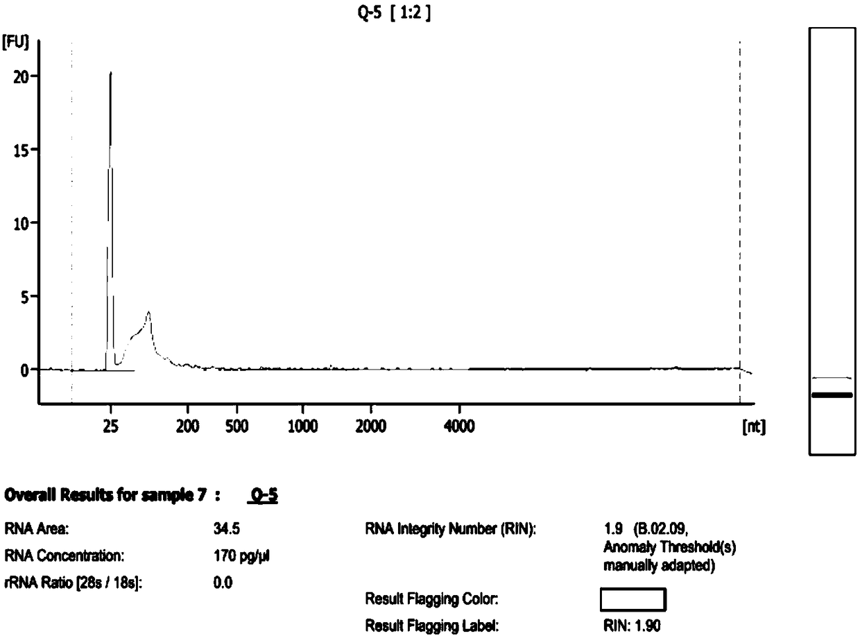
[0090] Exosome RNA obtained in Example 2 (sample number is sample7: Q-5)

[0091] CD63, TSG101, Calnexin (purchased from Abcam)

[0092] Marker: purchased from Thermo Scientific (#26617)

[0093] PVDF membrane: purchased from Immobilon-P (Cat No.IPVH00010)

[0094] Primary antibody diluent: purchased from Beyotime (P0023A)

[0095] HELA: Provided by the Institute of Basic Research, Chinese Academy of Medical Sciences

[0096] TBST: It is configured by 10×TBS plus 0.1% Tween-20

[0097] 10×TBS buffer:

[0098] Tris Base 24.2g

[0099] NaCl 80g

[0100] wxya 2 O 1000ml

[0101] TBST buffer:

[0102] 10×TBS 100ml

[0103] wxya 2 O 900ml

[0104] Tween-20 1ml

[0105] 2. Method -

[0106] (1) RNA quality inspection

[0107] BGI was entrusted to use the Agilent 2100 bioanalyzer for RNA quality in...


PUM

PropertyMeasurementUnit
diameteraaaaaaaaaa
diameteraaaaaaaaaa

Abstract

The invention relates to a method for separating and preparing exosomes, belonging to the field of biotechnology. The method for separating and preparing exosome comprise that following steps: 1) removing cellular impurities by centrifugation; 2) centrifuging to remove cellular debris and large protein impurities; 3) transferring that supernatant into a new centrifugal tube, centrifuging to discard the precipitate and leave the supernatant; 4) filtering that supernatant and transferring; 5) mixing that reagent XBP and the sample, transferring the reagent XBP into a membrane adsorption column,centrifuging, and discarding the liquid at the bottom of the tube; 6) centrifuging to remove liquid remaining on that membrane; 7) adding reagent XWP, centrifuging, discarding that liquid at the bottom of the tube, and changing the column into a new centrifugal tube; and 8) adding that reagent XE to the column, centrifuging and eluting the exocrine body to obtain the liquid containing the exocrinebody. The RNA purity of the extracted exocrine body is improved, the impurity is less, the biological sample volume of the initially extracted exocrine body can be reduced, and the yield of the circulating exocrine body can be improved.

Description

technical field [0001] The invention relates to a method for separating and preparing exosomes, belonging to the field of biotechnology. Background technique [0002] Multiple myeloma (MM) is a common hematologic tumor in middle-aged and elderly people, accounting for about 1% of all malignant tumors and 10% of hematologic malignancies. As my country gradually enters the aging process, its incidence has shown an obvious upward trend in recent years. The understanding of the pathogenesis and biological behavior of MM has gradually deepened, and a variety of effective therapeutic drugs have entered clinical application. MM has changed from a tumor with a low response rate to a disease that is treatable and controllable with various treatments. However, MM is still an incurable malignant tumor, and almost all MM will relapse and eventually fail to treat. [0003] At present, the most important and basic diagnostic and prognostic evaluation methods for MM are based on bone mar...

Claims


Application Information

Patent Timeline
Patent Type & Authority Applications(China)
IPC IPC(8): C12N5/09
CPCC12N5/0694C12N2509/00
Inventor 郝牧宫达森魏晓晶邱录贵
Owner INST OF HEMATOLOGY & BLOOD DISEASES HOSPITAL CHINESE ACADEMY OF MEDICAL SCI & PEKING UNION MEDICAL COLLEGE
Why Eureka
Try Eureka