G-quadruplex fluorescent dye and method based on highly conserved region G-quadruplex structure of HCV genome

A fluorescent dye and quadruplex technology, which is applied in the field of G-quadruplex fluorescent dyes based on the G-quadruplex structure in the highly conserved region of the HCV genome, can solve the problem of unfavorable G-quadruplex detection and the inability to truly reflect HCV Infection and replication, low transmembrane efficiency, etc.

Active Publication Date: 2022-04-08
HUNAN UNIV
View PDF1 Cites 0 Cited by
  • Summary
  • Abstract
  • Description
  • Claims
  • Application Information

AI Technical Summary

Problems solved by technology

However, commercial anti-HCV diagnostic reagents can not truly reflect the infection and replication of HCV in the body, although they can achieve specific diagnosis by binding to the surface antigen of the virus.
[0005] As a common G-quadruplex dye, Thioflavin T is widely used in nucleic acid detection systems, but because Thioflavin T itself is easily soluble in water, its transmembrane efficiency is low, which is not conducive to cell imaging technology. For the detection of intracellular G-quadruplex

Method used


Image

  • G-quadruplex fluorescent dye and method based on highly conserved region G-quadruplex structure of HCV genome
  • G-quadruplex fluorescent dye and method based on highly conserved region G-quadruplex structure of HCV genome
  • G-quadruplex fluorescent dye and method based on highly conserved region G-quadruplex structure of HCV genome

Examples


Embodiment 1

[0028] The structure of the G-quadruplex fluorescent dye is shown in formula (I):

[0029]

Embodiment 2

[0031] The preparation method of the G-quadruplex fluorescent dye described in embodiment 1 comprises the following steps (the synthesis route is shown in figure 2 ):

[0032] (1) Synthesis of intermediate 1: 2-methylbenzothiazol-6-amine (10mM, 1.64g) was used as a raw material, DMF was used as a solvent, methyl iodide (50mM, 7.09g) was added, and refluxed at 80 degrees for 18 hours, After the reaction was cooled to room temperature, the reaction mixture was suction-filtered, rinsed with cold ether (50-150 mL), the precipitate was collected, and vacuum-dried to obtain the product 2-amino-3,6-dimethylbenzothiazol-3-ium (intermediate 1, 2.44g, yield 80%);

[0033] (2) Synthesis of Intermediate 2: Intermediate 1 (5mM, 1.53g), potassium hydroxide aqueous solution (10M, 29g), ethylene glycol 6-12mL was added to a 100mL round-bottomed flask to react for 18h at reflux temperature, and the reaction was cooled to After room temperature, the mixture was poured into a beaker containin...

Embodiment 3

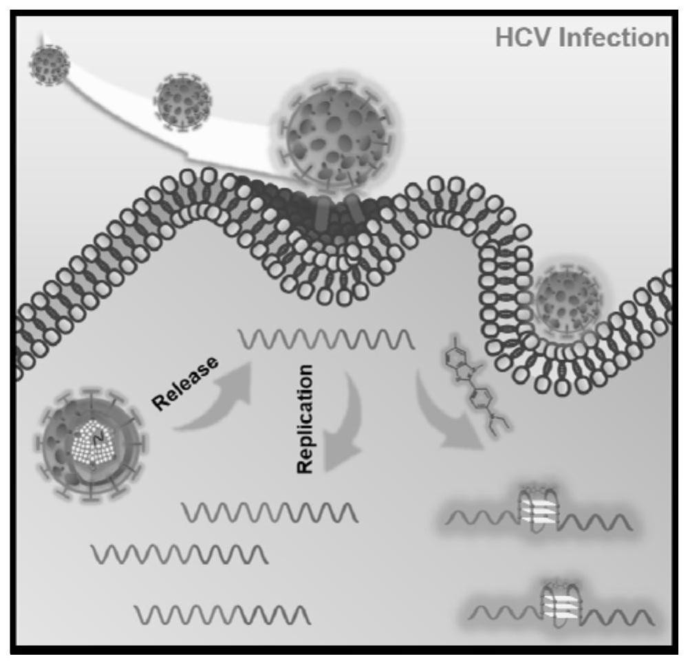
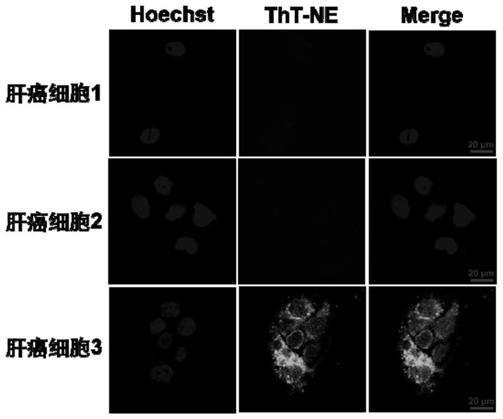
[0036] The detection method based on the HCV infection of the G-quadruplex fluorescent dye described in Example 1 is: preferably contain the live cell system (cell sample number is 5 * 10) that replicates active HCV infection 4 each, the reaction system was 100 μL) were co-incubated with ThT-NE, washed twice with phosphate-buffered saline, 5 min each time, and used a confocal laser fluorescence microscope (the parameter 403nm channel was used for living cell nuclear dye Hoechst imaging, and the 488nm channel was used for live cell nuclear dye Hoechst imaging). Cellular HCV RNA combined with ThT-NE imaging) is detected, the incubation time is 10-30 min, and the reaction temperature is 20-40°C.

[0037] Wherein, the dosage of the ThT-NE dye is 6.97-34.75ng; the excitation wavelength of the selected fluorescence microscope is blue light 460-488nm.

[0038] Test results:

[0039] The degree of infection is reflected by the imaging detection of actively replicating HCV-infected ce...


PUM


Abstract

The invention discloses a G-quadruplex fluorescent dye based on the G-quadruplex structure of the highly conserved region of the HCV genome, the structure of which is shown in formula (I). Based on the topological structure of the functional nucleic acid in the highly conserved region of the HCV genome, that is, the G-quadruplex structure, the present invention proposes a novel HCV detection idea, that is, the synthesized novel G-quadruplex dye can quickly enter cells and interact with cells The G-quadruplex structure in the HCV genome specifically binds to generate a highly sensitive fluorescent signal, and realizes the detection of HCV infection in living cells.

Description

technical field [0001] The invention relates to a G-quadruplex fluorescent dye based on the G-quadruplex structure of the highly conserved region of the HCV genome, a synthesis method and a detection method for live cell HCV (hepatitis C virus) infection. Background technique [0002] More than 170 million people worldwide are currently infected with the hepatitis C virus, HCV. As a major human pathogen, HCV belongs to the Flaviviridae family and is a positive-sense single-stranded RNA virus that produces a large number of daughters by inhibiting the host cell and replicating and expressing its own genetic material in the cytoplasmic environment of the host cell. Generation RNA and progeny virus. Once the human body is infected, it is very likely to develop diseases such as chronic hepatitis, fatty liver, liver fibrosis, liver cirrhosis and even liver cancer. What's more serious is that due to the inability to clarify the pathogenesis of hepatitis C, there is currently no ...

Claims


Application Information

Patent Timeline
Patent Type & Authority Patents(China)
IPC IPC(8): C07D277/66C09B57/00C09K11/06G01N21/64
CPCG01N21/6428C09K11/06C07D277/66C09B57/00C09K2211/1037C09K2211/1007G01N2021/6439
Inventor 聂舟骆星宇冯光富
Owner HUNAN UNIV
Why Eureka
Try Eureka