Bone-cartilage bidirectional function bracket based on cell 3D printing, and preparation method thereof

A 3D printing and cartilage technology, applied in medical science, prosthesis, tissue regeneration, etc., can solve the problem of single function of the scaffold, achieve clear chemical structure, convenient purification, and beneficial to tissue regeneration

Inactive Publication Date: 2019-04-26
STOMATOLOGY AFFILIATED STOMATOLOGY HOSPITAL OF GUANGZHOU MEDICAL UNIV
View PDF5 Cites 6 Cited by
  • Summary
  • Abstract
  • Description
  • Claims
  • Application Information

AI Technical Summary

Problems solved by technology

[0006] In order to overcome the defect of the single function of the scaffold described in the above prior art, the prese

Method used


Image

  • Bone-cartilage bidirectional function bracket based on cell 3D printing, and preparation method thereof
  • Bone-cartilage bidirectional function bracket based on cell 3D printing, and preparation method thereof
  • Bone-cartilage bidirectional function bracket based on cell 3D printing, and preparation method thereof

Examples


Embodiment 1

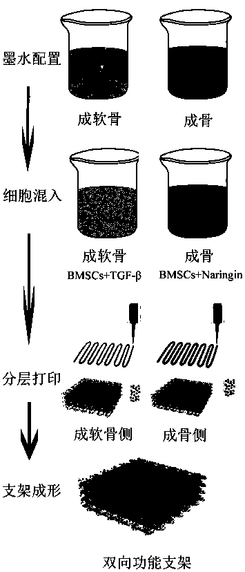
[0038] A method for preparing a bone-cartilage bidirectional functional scaffold, comprising the steps of:

[0039] S1. Configure bone-forming ink matrix and chondrogenic ink matrix, and prepare them after disinfection; wherein, the bone-forming ink matrix includes the following mass percentages: 10% mesoporous bioglass powder, 5% sodium alginate, 5% gelatin, and the rest for water;

[0040]The chondrogenic ink matrix includes the following composition in mass percentage: 10% sodium alginate, 2.5% gelatin, and the balance is water.

[0041] S2. Add bone marrow mesenchymal stem cells (BMSCs) and naringin (Naringin) into the matrix of bone-forming ink to obtain bone-forming ink; in the bone-forming ink, the concentration of BMSCs is 5×10 6 / mL, the concentration of naringin is 1×10 -5 mol / L.

[0042] BMSCs and transforming growth factor β (TGF-β) (1 × 10 -9 mol / L) was added into the matrix of the chondrogenic ink to obtain the chondrogenic ink. In the chondrogenic ink, th...

Embodiment 2

[0047] A method for preparing a bone-cartilage bidirectional functional scaffold, comprising the steps of:

[0048] S1. Configure bone-forming ink matrix and chondrogenic ink matrix, and prepare them after disinfection; wherein, the bone-forming ink matrix includes the following components by mass percentage: 5% mesoporous bioglass powder, 10% sodium alginate, 2.5% gelatin, and the balance for water;

[0049] The chondrogenic ink matrix includes the following composition in mass percentage: 10% sodium alginate, 5% gelatin, and the balance is water.

[0050] S2. Add bone marrow mesenchymal stem cells (BMSCs) and naringin (Naringin) into the matrix of bone-forming ink to obtain bone-forming ink; in the bone-forming ink, the concentration of BMSCs is 5×10 6 / mL, the concentration of naringin is 1×10 -5 mol / L.

[0051] Add BMSCs and transforming growth factor β (TGF-β) into the matrix of chondrogenic ink to obtain chondrogenic ink. In the chondrogenic ink, the concentration ...

Embodiment 3

[0056] A method for preparing a bone-cartilage bidirectional functional scaffold, comprising the steps of:

[0057] S1. Configure bone-forming ink matrix and chondrogenic ink matrix, and prepare them after disinfection; wherein, the bone-forming ink matrix includes the following components by mass percentage: 5% mesoporous bioglass powder, 10% sodium alginate, 2.5% gelatin, and the balance for water;

[0058] The chondrogenic ink matrix includes the following composition in mass percentage: 10% sodium alginate, 5% gelatin, and the balance is water.

[0059] S2. Add bone marrow mesenchymal stem cells (BMSCs) and naringin (Naringin) into the matrix of bone-forming ink to obtain bone-forming ink; in the bone-forming ink, the concentration of BMSCs is 5×10 6 / mL, the concentration of naringin is 1×10 -4 mol / L.

[0060] Add BMSCs and transforming growth factor β (TGF-β) into the matrix of chondrogenic ink to obtain chondrogenic ink. In the chondrogenic ink, the concentration ...


PUM

PropertyMeasurementUnit
Concentrationaaaaaaaaaa

Abstract

The invention discloses a bone-cartilage bidirectional function bracket based on cell 3D printing, and a preparation method thereof. The preparation method for the bone-cartilage bidirectional function bracket based on cell 3D printing comprises the following steps: S1: respectively preparing an osteogenesis ink substrate and a chondrogenesis ink substrate; S2: adding mesenchymal stem cells and naringin into the osteogenesis ink substrate to obtain osteogenesis ink, and adding the mesenchymal stem cells and a transforming growth factor beta into the chondrogenesis ink substrate to obtain chondrogenesis ink; S3: respectively printing the osteogenesis ink and the chondrogenesis ink to obtain a bracket, wherein the bracket comprises an osteogenesis side and a chondrogenesis side; S4: after the bracket is subjected to crosslinking treatment, obtaining the bone-cartilage bidirectional function bracket. According to the bone-cartilage bidirectional function bracket prepared with the preparation method, the combined regeneration of bone-cartilage can be simultaneously realized, personalized customization is realized, the bone-cartilage bidirectional function bracket is favorable for the tissue regeneration of joints, and therefore, damaged joints can be more favorably repaired.

Description

technical field [0001] The present invention relates to the field of tissue engineering, more specifically, to a bone-cartilage bidirectional functional scaffold based on cell 3D printing and a preparation method thereof. Background technique [0002] Bone tissue has the function of self-repair and regeneration and reconstruction, but the large-area bone defect caused by trauma, tumor, infection, osteoporosis caused by aging population, etc., is limited only by the self-repair ability of bone, especially in joints. Cartilage, which cannot heal itself after injury. At present, the commonly used clinical treatment methods mainly include microfracture, autologous chondrocyte transplantation, and artificial joint replacement. These repair methods have their own shortcomings. The rise of tissue engineering provides a new direction for the study of cartilage defect repair. [0003] Rapid prototyping technology is also called 3D printing technology. Low temperature rapid prototy...

Claims


Application Information

Patent Timeline
IPC IPC(8): A61L27/22A61L27/20A61L27/10A61L27/38A61L27/50A61L27/54A61L27/58
CPCA61L27/10A61L27/20A61L27/222A61L27/3834A61L27/50A61L27/54A61L27/58A61L2300/232A61L2300/412A61L2300/414A61L2430/24C08L5/04
Inventor 郭吕华武敬文任文魏娟梁婷婷
Owner STOMATOLOGY AFFILIATED STOMATOLOGY HOSPITAL OF GUANGZHOU MEDICAL UNIV
Why Eureka
Try Eureka