Tumor cell heterogeneic transplantation zebrafish model, construction method and application thereof

A technology of xenotransplantation and tumor cells, which is applied in the field of zebrafish model of gastric cancer xenografting, can solve the problems that PDX models cannot meet

Inactive Publication Date: 2019-05-14
NANJING EMORY BIOTECH CO LTD
View PDF5 Cites 1 Cited by
  • Summary
  • Abstract
  • Description
  • Claims
  • Application Information

AI Technical Summary

Problems solved by technology

At present, mice are the most commonly used tumor PDX model animals. However, since the time for mouse tumor inoculation, tumor formation and drug efficacy evaluation is usually 3 months, and the survival period of many patients is less than 3 months, the existing PDX The model cannot meet the important needs of clinical real-time guidance of individualized medicine

Method used


Image

  • Tumor cell heterogeneic transplantation zebrafish model, construction method and application thereof
  • Tumor cell heterogeneic transplantation zebrafish model, construction method and application thereof
  • Tumor cell heterogeneic transplantation zebrafish model, construction method and application thereof

Examples


Embodiment 1

[0049] Example 1: Construction of the patient-derived gastric cancer cell xenograft zebrafish model of the present invention

[0050] 1. Isolation of Primary Cells from Gastric Cancer Tissue

[0051] Surgical specimens of gastric cancer from patient-derived clinical tissue biopsies were placed in normal saline, blood clots, necrotic tissue, fat, and connective tissue on the tumor tissue surface were removed under aseptic conditions, and the tissue was cut into pieces with sterilized ophthalmic scissors. Wash twice with sterile phosphate buffer (pH 7.4), add a small amount of phosphate buffer, and repeatedly cut the tissue with elbow ophthalmic scissors until the tissue becomes a paste, about 1mm 3 size. Add 0.25% trypsin, digest at 37°C for 10 minutes, and centrifuge to remove the trypsin after observing that the tissue pieces are completely dissociated. The cells were resuspended in RPMI-1640 medium containing 10% FBS (fetal bovine serum).

[0052] 2. Staining of Primary C...

Embodiment 2

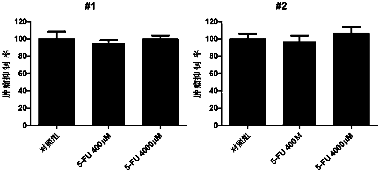
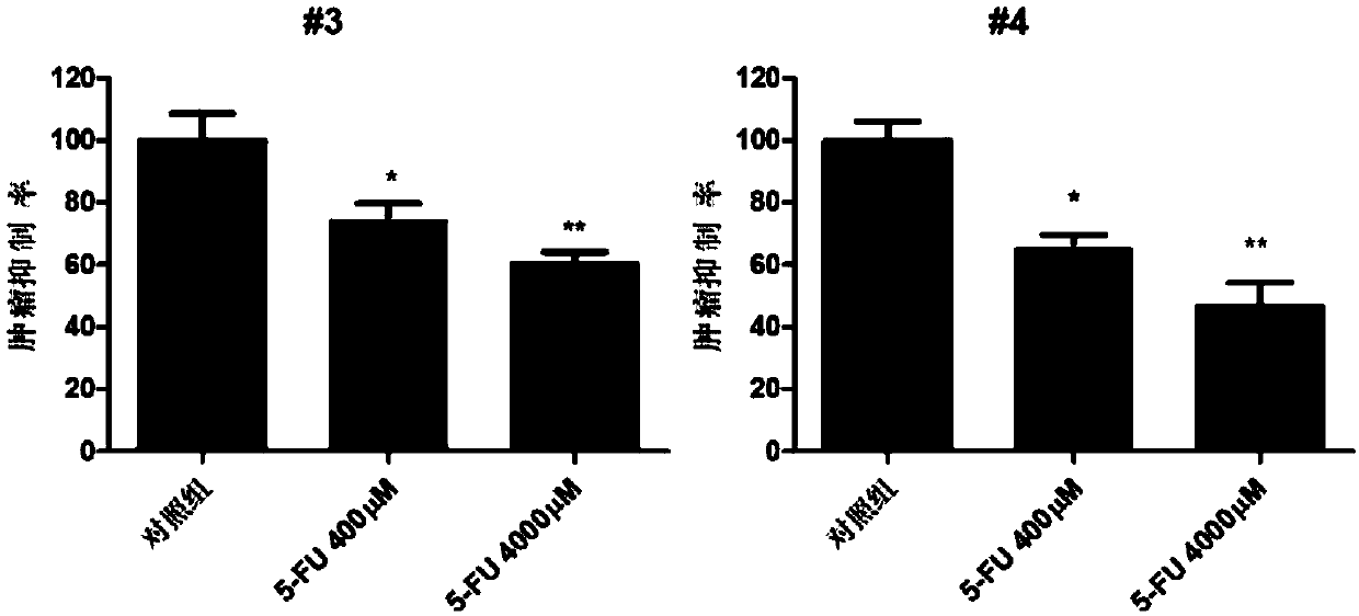
[0059] Example 2: 4 patient-derived xenograft zebrafish models are used to evaluate the clinical anticancer effect of 5-FU

[0060] 1. Determination of safe dose

[0061] Zebrafish embryos two days after fertilization were treated (soaked) with different concentrations of 5-FU for three consecutive days, and the highest concentration of 5-FU within the safe range of embryos was determined to be 4000 μM.

[0062] 2. Drug Treatment of Zebrafish Embryos

[0063] 4000 μM and 400 μM 5-FU were selected to act (soak) the zebrafish embryo models prepared by the method in Example 1 and injected with gastric cancer primary cells from different patients for three consecutive days, and 0.1% DMSO was used as a solvent control.

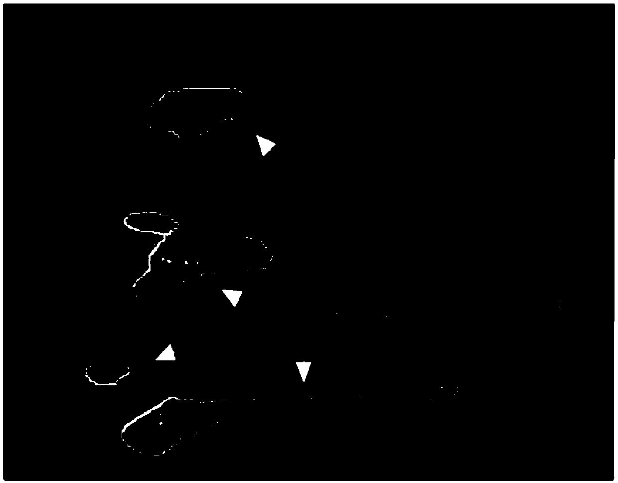
[0064] 3. Observation of anti-tumor effect by fluorescence microscope

[0065] Observe and compare the proliferation and spread of red patient-derived cells in the treated zebrafish embryos. The red cells were photographed under a fluorescence microscope, and th...

Embodiment 3

[0068] Example 3: Two xenograft zebrafish models of human gastric cancer cell lines (SGC-7901 and AGS) were used to evaluate the anticancer effect of 5-FU

[0069] 1. Drug Treatment of Zebrafish Embryos

[0070] Select 4000 μM and 400 μM 5-FU to act on (soak) zebrafish embryos (constructed according to the method in Example 1) that have been injected with gastric cancer cell lines (SGC-7901 and AGS) for three consecutive days, and use 0.1% DMSO as a solvent control.

[0071] 2. Observation of anti-tumor effect by fluorescence microscope

[0072] Observe and compare the proliferation and spread of red patient-derived cells in the treated zebrafish embryos. The red cells were photographed under a fluorescence microscope, and the red fluorescence intensity was quantified with Image Pro Plus software to calculate the anti-tumor effect of 5-FU. fluorescence intensity)*100% (see Figure 4 ).

[0073] The results of the xenograft zebrafish model of human gastric cancer cell line...


PUM


Abstract

The invention provides a tumor cell heterogeneic transplantation zebrafish model, a construction method and application thereof. By implanting primary cells separated from a tumor tissue of a patientinto a zebrafish, the tumor heterogeneic transplantation model derived from the patient is obtained. According to the established tumor heterogeneic transplantation model, pathological characteristicsof human gastric cancer tissue in clinic are preserved, higher clinical correlation is achieved, the established tumor heterogeneic transplantation model can be used for systematically studying the mechanism of tumor proliferation, metastasis, diffusion and drug resistance, and tumor therapeutic drugs are effectively screened.

Description

technical field [0001] The invention relates to the field of biomedicine, in particular to a patient-derived zebrafish model of gastric cancer xenograft, its construction method and application. Background technique [0002] Tumor diseases have become a major public health problem all over the world. The most common tumors are lung cancer, gastric cancer, and breast cancer. [0003] Among them, gastric cancer is one of the most common malignant tumors of the digestive system in the world, with the highest incidence in East Asia. According to the "Global Cancer Report 2014" released by the World Health Organization (WHO), the number of new gastric cancer cases and deaths in China accounted for more than 40% of the world's total in 2012. In 2016, the 2015 China Cancer Statistics Report published in the authoritative journal "CA Cancer JClin" sponsored by the American Cancer Society (ACS) showed that there were 679,100 new cases of gastric cancer in China in 2015, and the numb...

Claims


Application Information

Patent Timeline
Patent Type & Authority Applications(China)
IPC IPC(8): A01K67/027
Inventor 何明芳檀爱民王瑞雪李建英
Owner NANJING EMORY BIOTECH CO LTD
Why Eureka
Try Eureka