A high-throughput functional gene screening method and system for cell phenotypic image quantitative analysis

A technology for functional gene and quantitative analysis, applied in image analysis, material analysis, sequence analysis, etc., can solve problems such as low throughput and inability to accurately quantify cell phenotypes, achieving good repeatability, fast identification, and reduced workload Effect

Active Publication Date: 2019-05-28
HUAZHONG UNIV OF SCI & TECH
View PDF14 Cites 9 Cited by
  • Summary
  • Abstract
  • Description
  • Claims
  • Application Information

AI Technical Summary

Problems solved by technology

[0005] The present invention solves the technical problem of low flux in cell screening technology in the prior art and cannot accurately quantify cell phenotype, and provides a high-throughput functional gene screening method, thereby greatly improving screening efficiency and improving results The degree of accuracy, and the functional genes can be sorted and classified according to the time point and degree of their influence on the cell phenotype, so as to build a gene interaction network

Method used


Image

  • A high-throughput functional gene screening method and system for cell phenotypic image quantitative analysis
  • A high-throughput functional gene screening method and system for cell phenotypic image quantitative analysis
  • A high-throughput functional gene screening method and system for cell phenotypic image quantitative analysis

Examples


Embodiment 1

[0042] The 103 phosphorylation regulator genes in the iEKPD database were screened to see whether they were related to autophagy.

[0043] 1. Generate a training set: import GFP-Atg8 into yeast cells to express and produce green fluorescence, use FM4-64 to mark vacuoles to produce red fluorescence, and the conditions for phenotype generation include nitrogen reduction and rapamycin induction (rapamycin induction) Wait. The cells were non-autophagy phenotype cells at 0 hours, and the cells underwent autophagy after 2 hours under reduced nitrogen conditions. Generate large numbers of images of phenotyped and non-phenotyped cells using fully automated fluorescence microscopy.

[0044] 2. Image processing: enhance the fluorescence signal by enhancing the contrast and deconvolution and denoising. Deconvolution and denoising: use PSF (point spread function) of different sizes to restore blurred images, analyze and reconstruct PSF, so as to improve the restored image.

[0045] 3. ...

Embodiment 2

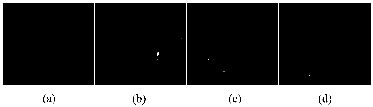
[0060] figure 1 (a) Cell phenotype at 0h after atg1 knockout under nitrogen reduction condition, no autophagy at this time; figure 1 (b) Cell phenotype at 2 hours after atg1 knockout under nitrogen reduction condition, at which point autophagy did not occur; figure 1 (c) is the cell phenotype at 0h after SNF1 is knocked out under nitrogen reduction condition, and there is no autophagy at this time; figure 1(d) Cell phenotype at 2 h after SNF1 knockout under nitrogen reduction condition, at which time autophagy occurs. Such as figure 1 As shown in (d), GFP-Atg8 enters FM4-64-labeled vacuoles during autophagy; as figure 1 (a), figure 1 (b) and figure 1 As shown in (c), GFP-Atg8 is mainly localized outside the vacuole when cells are not autophagy. We can teach computers to recognize this phenotypic difference to distinguish autophagic from non-autophagic cells.

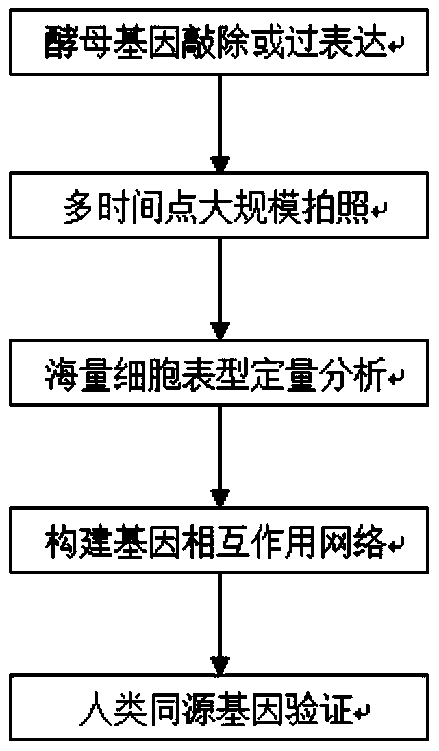
[0061] figure 2 It is a flowchart of a high-throughput gene screening method based on quantitative analysis o...


PUM


Abstract

The invention discloses a high-throughput functional gene screening method and system for cell phenotypic image quantitative analysis, and belongs to the technical field of gene screening. The methodcomprises the steps of using a full-automatic fluorescence microscope to shoot a cell image of the phenotype to be screened and a cell image without the phenotype to be screened; respectively converting the images into a to-be-screened phenotype and a black-white binary image without the to-be-screened phenotype; then segmenting into an image containing a single phenotypic cell to be screened andan image containing a single phenotypic cell without being screened; taking the corresponding part of the image containing the single to-be-screened phenotypic cell in the cell image of the to-be-screened phenotypic cell as a positive training set, and taking the corresponding part of the image containing the single to-be-screened phenotypic cell in the cell image of the to-be-screened phenotypiccell as a negative training set to obtain a final model capable of identifying the cell phenotypic; and carrying out final model identification on the image of the knockout or overexpression cell of the to-be-screened gene to obtain the relevance between the to-be-screened gene and the phenotype. The method improves the screening efficiency and accuracy.

Description

technical field [0001] The present invention relates to the technical field of gene screening, more specifically, to a high-throughput functional gene screening method and system for quantitative analysis of cell phenotype images. Background technique [0002] Many experimentalists are committed to identifying functional genes related to specific cell behaviors and constructing the link between genes and phenotypes, which will help humans interpret genes, understand diseases, and develop drugs. For example, the abnormality of autophagy is closely related to the occurrence of various diseases including cancer. Japanese scientist Yoshinori Ohsumi discovered the key gene regulating autophagy through yeast gene screening experiments. Because of his contribution to autophagy, he was awarded the Nobel Prize in Medicine in 2016. Yoshinori Ohsumi disclosed a technique using SDS-PAGE to detect GFP-ATG8 cleavage. This technique uses the difference in mobility between GFP-ATG8 and GFP...

Claims


Application Information

Patent Timeline
Patent Type & Authority Applications(China)
IPC IPC(8): G06K9/00G06K9/34G06T7/60G16B30/00G01N21/84
Inventor 薛宇宁万山郭亚萍
Owner HUAZHONG UNIV OF SCI & TECH
Why Eureka
Try Eureka