3D printed adipose tissue

A 3D printing, adipose tissue technology, applied in tissue culture, bone/connective tissue cells, drug combination, etc., can solve the problems of limited application of adipose tissue treatment, limited quantity, difficult transplantation of soft tissue, etc., to achieve the function of cell secretion. Effect

Inactive Publication Date: 2019-07-02
SHENZHEN INST OF ADVANCED TECH
View PDF4 Cites 1 Cited by
  • Summary
  • Abstract
  • Description
  • Claims
  • Application Information

AI Technical Summary

Problems solved by technology

However, pure brown adipose tissue transplantation, due to the limited number of donor cells and tissues, the soft tissue is not easy to transplant, and the limitations of clinical application, limits the application of adipose tissue in the treatment of related diseases. Therefore, combined with 3D printing technolog

Method used


Image

  • 3D printed adipose tissue
  • 3D printed adipose tissue
  • 3D printed adipose tissue

Examples


Embodiment 1

[0042] Embodiment 1 Configuration of mixed culture medium and adipocyte induction medium

[0043] The composition of the mixed culture medium is: DMEM F12+10% FBS+1% streptomycin+1% penicillin. The mixed culture solution was sterilized by 0.22 μm filter at 37 °C, 5% CO 2 Equilibrate in the incubator for 2-4 hours.

[0044]Adipocyte induction medium component I: MEM / F12+10%FBS+streptomycin+penicillin+5ug / mL insulin+1nM 3,3',5-triiodo-L-thyronine (T3); 0.22μm filter After sterilization at 37 °C, 5% CO 2 Equilibrate in the incubator for 2-4 hours.

[0045] Adipocyte induction medium component II: MEM / F12+10%FBS+streptomycin+penicillin+5ug / mL insulin+1nM3,3',5-T triiodo-L-thyronine (T3)+125uM indole Methacin + 0.5mM 3-isobutyl-1-methylxanthine (IBMX) + 0.5uM rosiglitazone; 0.22μm filter sterilization at 37°C, 5% CO 2 Equilibrate in the incubator for 2-4 hours.

Embodiment 2

[0046] Example 2 Obtaining brown adipocytes

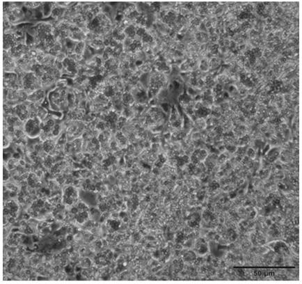
[0047] Take out the mouse scapular fat under aseptic conditions, carefully remove the surrounding white adipose tissue, wash 3 times with PBS (containing 1% penicillin and streptomycin), remove blood clots and mucus on the surface of the tissue, put it into a petri dish, and place the Cut up the adipose tissue and add collagenase D digestion mixture (1.5U / mL collagenase D+2.4u / mL DipaseⅡ+25mL PBS+10mM CaCl 2 ) 10ml, mix well, place in a water bath at 37°C for 60min, shake and digest; centrifuge at 700g for 10min, the adipocytes are located at the bottom of the centrifuge tube; after pouring off the supernatant, add 10ML of medium to resuspend the cells at the bottom, and repeatedly pipette to mix After uniformity, filter through a 70-mesh filter to remove undigested tissues, and collect the filtrate; obtain a crude mixture of adipocytes and other impurities.

[0048] Isolation of precursor cells in adipose tissue by differential a...

Embodiment 3

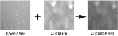
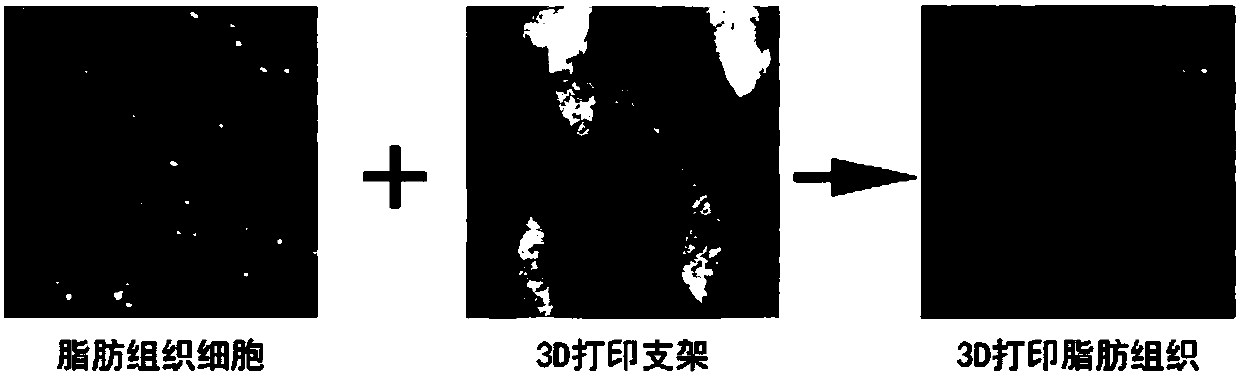
[0049] Example 3 Preparation of 3D printed brown adipose tissue

[0050] 1. Design and preparation of biocompatible scaffolds for 3D printed brown adipose tissue

[0051] (1) Combining with computer aided design (CAD), three-dimensional printing technology is used to construct the bracket, in which the shape, composition and internal structure are well designable;

[0052] (2) The shape of the three-dimensional adipose tissue is designed to be similar to the shape of adipose tissue, and the internal structure is guaranteed to have a certain pore structure;

[0053] (3) The compatibility of gelatin (10%) was selected as the printing material of the biocompatible scaffold for printing.

[0054] (4) Internal structural parameters of the constructed biocompatible scaffold: pore size (R) = 0.2mm; line stacking angle: 90°

[0055] (5) Store the prepared biocompatible scaffold at -80°C for future use.

[0056] 2. Preparation of 3D artificial adipose tissue

[0057] The preadipocy...


PUM


Abstract

The present invention relates to a 3D printed adipose tissue and particularly discloses the artificial adipose tissue. The artificial adipose tissue comprises a biocompatible stent, adipose cells anda mixed culture solution. Through a 3D bioprinting technique, the biocompatible stent and the adipose tissue are prepared into the biologically active artificial adipose tissue. The mixed culture solution is used for culture and preservation of the adipose cells and tissue. The biocompatible stent is prepared using the 3D printing technique. The artificial adipose tissue can be used as a pathophysiological mechanism research and therapeutic tool for adipose-related endocrine and metabolic diseases.

Description

technical field [0001] The invention relates to the field of biotechnology, in particular to adipose tissue prepared by 3D printing technology. Background technique [0002] Adipose tissue is composed of fat cells separated into lobules by a thin layer of loose connective tissue. Adipose tissue is not just a simple energy storage organ, its more important function is to participate in the pathological and physiological processes of various diseases through the endocrine system. Adipose tissue is divided into white adipose tissue and brown adipose tissue. White adipose tissue is composed of a large number of unicellular adipocytes, mainly distributed in subcutaneous, mesenteric and other parts. It is the energy bank in the body, and has the functions of storing fat, maintaining body temperature, regulating fat metabolism, and supporting protection. Brown fat is characterized by rich capillaries, in which adipocytes contain many fat droplets and are rich in mitochondria. Br...

Claims


Application Information

Patent Timeline
IPC IPC(8): C12N5/077A61K49/00A61P3/00A61P15/00A61P3/04A61P3/10A61P9/12A61P9/10A61P3/06
CPCA61K35/35A61K49/0008C12N5/0653C12N2533/54C12N2533/70C12N2533/72C12N2533/74C12N2533/76C12N2533/80
Inventor 张键黄晨赵华山汪宝蓓陈指龙薛丽杨雅莉
Owner SHENZHEN INST OF ADVANCED TECH
Why Eureka
Try Eureka