Preparation of rabbit anti-human TRK protein monoclonal antibody and immunohistochemical application thereof

A technology of monoclonal antibody and immunohistochemistry, which is applied in the field of immunohistochemistry, rabbit monoclonal antibody against human TRK protein, can solve the problem of rare TRK antibody, and achieve the effect of improving specificity and reliability

Inactive Publication Date: 2019-07-19
广东三九脑科医院 +1
View PDF3 Cites 0 Cited by
  • Summary
  • Abstract
  • Description
  • Claims
  • Application Information

AI Technical Summary

Problems solved by technology

Since the gene inhibitory drug has just been launched, there is no TRK antibody independently developed in China, and commercialized TRK antibodies are rare in the world, and they are only used in the field of basic research at present, and the

Method used


Image

  • Preparation of rabbit anti-human TRK protein monoclonal antibody and immunohistochemical application thereof
  • Preparation of rabbit anti-human TRK protein monoclonal antibody and immunohistochemical application thereof
  • Preparation of rabbit anti-human TRK protein monoclonal antibody and immunohistochemical application thereof

Examples


Embodiment 1

[0023] Embodiment 1 Preparation of anti-TRK monoclonal antibody

[0024] 1. Preparation of immune source: According to the sequence information of the three subtypes of NTRK gene NTRK1 (NM_002529.3), NTRK2 (NM_001018064.2) and NTRK3 (NM_001012338.2), order and synthesize human TRK universal polypeptide with BSA sequence at the N-terminal , for immunization of experimental animals and ELISA screening of positive clones.

[0025] 2. Blood collection before immunization: select purebred New Zealand white rabbits, and take blood from ear veins as pre-immunization blood samples.

[0026] 3. Screening and preparation of monoclonal antibodies

[0027] 1. Animal immunization: The above synthetic human TRK universal polypeptide with BSA sequence at the N-terminal was fully mixed with an equal volume of Freund's complete adjuvant to prepare a fully emulsified immune source, and the purebred New Zealand white rabbit was first immunized by subcutaneous injection. The immunization dose i...

Embodiment 2

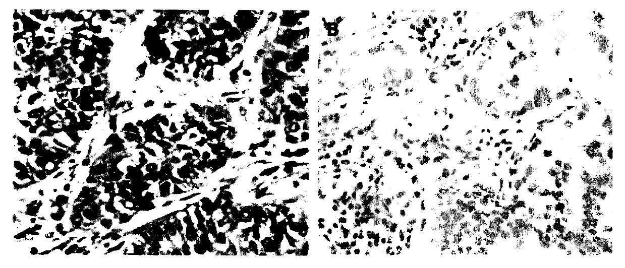
[0030] Example 2 Immunohistochemical experiments using the monoclonal antibody of the present invention as the primary antibody

[0031] 1. Sampling 24 different types of cancer tissues to make tissue microarrays, and slice them with a Leica RM2235 tissue slicer with a thickness of 4 μm;

[0032] 2. Use the Leica BondMax immunohistochemical automatic staining machine to carry out immunohistochemical staining test on the antibody of the present invention, using the dewaxing and hydration conditions that come with the machine. The specific steps are: incubate at 60°C for 30min, and wash with dewaxing solution (Leica). 3 times. For antigen retrieval, antigen retrieval solution 2 (ER2, Leica) was used and incubated at 100°C for 30 min. The antibody of the present invention was used as the primary antibody, diluted with antibody diluent (Leica) to a final concentration of 1.0 μg / ml, 150 μl. The antibody was incubated at room temperature for 30 min. Use 150 μl of matching seconda...

Embodiment 3

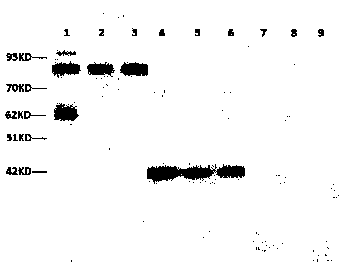
[0035] Embodiment 3 Specific detection of monoclonal antibody of the present invention

[0036] No positive band can be seen in the detection of untransfected 293T cells by using the antibody of the present invention.

[0037] The antibody of the present invention was used to detect the 96-well plate (Her-2) pre-coated with irrelevant antigens by ELISA, and the result was negative.


PUM

PropertyMeasurementUnit
Thicknessaaaaaaaaaa

Abstract

The invention discloses a rabbit anti-human TRK protein monoclonal antibody, can specifically recognize the TRK and/or TRK fusion protein expressed by cells, has stable performance and high titer, hasno cross-reaction with other proteins in the cell, and has certain medical basic research value. The invention also relates to the use of the above monoclonal antibody for the preparation of immunohistochemical detection tools for detecting TRK and/or TRK fusion protein. The application adopts an immunohistochemical method to detect TRK and/or TRK protein of tumor cells including lung cancer, thyroid cancer, melanoma, gastrointestinal cancer, colorectal carcinoma, soft tissue sarcoma, salivary gland, infant fibrosarcoma, appendix cancer, breast cancer, cholangiocarcinoma, pancreatic cancer, and the like. The monoclonal antibody provides a powerful scientific reference for the treatment of TRK kinase inhibitors, effectively reduces the cost of treatment, and has important application value.

Description

technical field [0001] The invention relates to the field of biotechnology, in particular to a rabbit monoclonal antibody against human TRK protein and the immunohistochemical application of the antibody. Background technique [0002] Tropomyosin-related kinase (TRK) is a type of nerve growth factor receptor involved in the regulation of cell communication and tumor growth, and NTRK is the gene encoding TRK. Gene NTRK includes NTRK1, NTRK2 and NTRK3, responsible for encoding the synthesis of TRK family proteins TRKA, TRKB and TRKC, respectively. TRK family proteins all have a homologous and conserved structural region, which includes 5 extracellular domains, 1 transmembrane domain and 1 intracellular kinase domain. Among the five extracellular domains, domains 1 and 3 are cysteine-rich regions, domain 2 is leucine-rich regions, and domains 4 and 5 are immunoglobulin-like structures. Domain 5 is the site for ligand-specific recognition. With the deepening of research, more...

Claims


Application Information

Patent Timeline
IPC IPC(8): C07K16/40G01N33/577G01N33/573G01N33/574
CPCC07K16/40G01N33/577G01N33/573G01N33/57492G01N2333/912
Inventor 李智李海南蔡林波李军李沛祥李雪方洁羽
Owner 广东三九脑科医院
Why Eureka
Try Eureka