Detection method for respiratory tract viruses

A detection method and respiratory technology, applied in the biological field, can solve the problems of high requirements for instruments and operating techniques, increased detection costs, time consumption and server space

Active Publication Date: 2019-08-06
INST OF PATHOGEN BIOLOGY CHINESE ACADEMY OF MEDICAL SCI
View PDF6 Cites 17 Cited by
  • Summary
  • Abstract
  • Description
  • Claims
  • Application Information

AI Technical Summary

Problems solved by technology

That is, in the process of next-generation sequencing, in order to increase the coverage of viral nucleic acid, the only way to increase the sequencing depth is to increase the amount of sequencing data for each sample. Such a result will generate a large number of reads (sequenced fragments) and genetic information. Hiseq sequencing can The amount of data reaches 200G, and Miseq sequencing can also reach the amount of data of 15G. However, when analyzing these data in bioinformatics, conventional methods process the data generated by samples, and it is found that more than 99% of them are host gene sequences, especially low-copy viruses. For samples, the number of virus reads is less than 0.01%, which leads to a direct increase in the cost of next-generation sequencing, which is not conducive to the application of next-generation sequencing of respiratory viruses in clinical samples. In addition, massive data analysis not only increases the detection cost but also consumes a lot of time and time. server space
In order to solve this problem, many researchers enrich the virus by ultracentrifugation. Although the flux of the virus has been improved to a certain extent, ultracentrifugation takes at least six hours, and the number of samples in each centrifugation is limited to 6. The requirements for instruments and operating techniques are very high, so this method is not suitable for high-throughput NGS detection of a large number of clinical samples

Method used


Image

  • Detection method for respiratory tract viruses
  • Detection method for respiratory tract viruses
  • Detection method for respiratory tract viruses

Examples


Embodiment 1

[0112] We take the virus detection of the respiratory samples of 4 cases of human lung diseases collected in June 2018 as an example to illustrate the specific embodiments of the present invention in sequence.

[0113] 1. Sample collection and storage conditions:

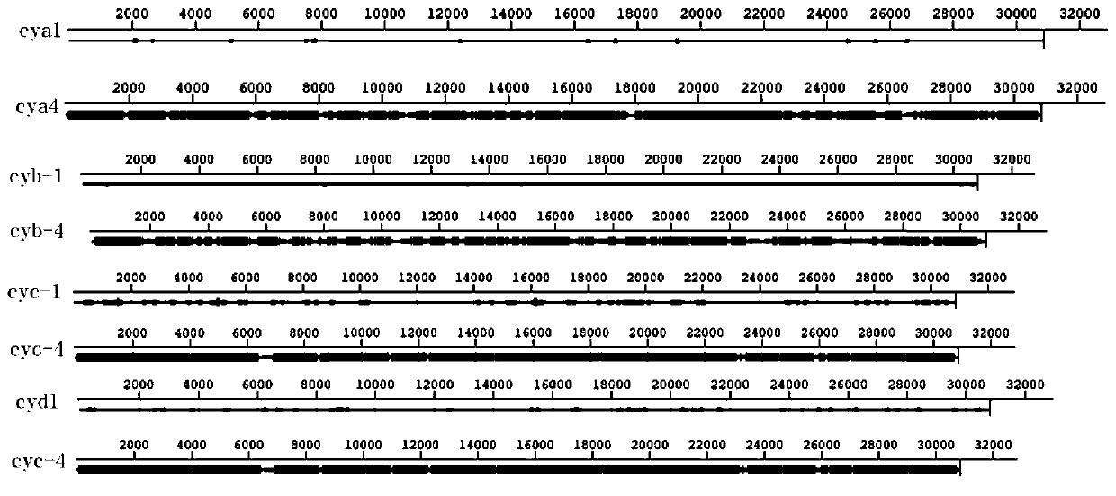
[0114] 1. First, take throat samples from inpatients diagnosed as COPD patients (cya), allergic patients (cyb), pulmonary hemorrhage patients (cyc) and lung cancer patients (cyd) and quickly store them in virus collection tubes (about 3ml) , oscillate for 30s. Other trace samples can also be stored in virus collection tubes. For the convenience of comparison, each sample is processed by two methods, and processed according to the method of the present invention. The samples are named cya1, cyb1, cyc1 and cyd1, and the samples processed by modern methods are named as cya2cyb2, cyc2 and cyd2 directly extract nucleic acid without going through the enrichment process of the method.

[0115] 2. The amount of sampling c...


PUM


Abstract

The invention provides a high-throughput detection method for efficient and rapid enrichment of respiratory tract viruses. The high-throughput detection and identification technology for efficient andrapid enrichment of the respiratory tract viruses includes the following four parts: enrichment of respiratory tract sample viruses, library building, high-throughput sequencing and biological information analysis. Enrichment of the respiratory tract sample viruses is a core part of the technology. Firstly, nucleic acids of hosts except viruses in the samples are removed by a biophysical method,and then sample viral nucleic acids are extracted. With the help of detection of an Agilent 2100 instrument, only 200 pg nucleic acids are used for building a library. The results of high-energy sequencing and biological information analysis show that the reads number of the viruses is increased from less than 0.1% with treatment by conventional methods to 84% or more, and the detection technologynot only can detect sample infected viruses quickly, increases the coverage rate (95-100% or more) of a whole genome sequence of the viruses, and greatly reduces the cost of sequencing.

Description

[0001] Technical field: [0002] The invention relates to the field of biotechnology, and relates to a detection method for respiratory viruses. [0003] Background technique: [0004] Viruses can cause a wide variety of human diseases. Respiratory tract infection is divided into upper respiratory tract infection and lower respiratory tract infection. Upper respiratory tract infection refers to the general term of acute inflammation between the nasal cavity and the throat, and is the most common infectious disease. Lower respiratory tract infection is the most common infectious disease, and the pathogen causing the infection must be clearly identified in order to select an effective treatment method. About 90% of upper respiratory tract infections are caused by viruses, and bacterial infections are often secondary to viral infections. This disease all can fall ill in four seasons, any age, spreads by droplet, fog drop that contains virus, or through contaminated utensils. O...

Claims


Application Information

Patent Timeline
Patent Type & Authority Applications(China)
IPC IPC(8): C12Q1/70C12Q1/6806C12Q1/6869C12R1/93
CPCC12Q1/6806C12Q1/6869C12Q1/701C12Q2521/107C12Q2531/113C12Q2525/191C12Q2535/122
Inventor 张婷杨帆金奇
Owner INST OF PATHOGEN BIOLOGY CHINESE ACADEMY OF MEDICAL SCI
Why Eureka
Try Eureka