Preparation of SERS substrate and application of SERS substrate to cancer detection

A substrate and detection system technology, applied in the field of nanomaterials, can solve the problems of long time consumption, high sensitivity, and low efficiency, and achieve the effects of short time consumption, enhanced SERS signal, and high sensitivity

Inactive Publication Date: 2019-08-13
WUHAN UNIV
View PDF4 Cites 5 Cited by
  • Summary
  • Abstract
  • Description
  • Claims
  • Application Information

AI Technical Summary

Problems solved by technology

At present, the use of SERS method to solve biological problems is a hot spot. The main reason is that traditional medical detection methods have the disadvantages of long time consumption and low efficiency, while SERS spectrum acquisition often only takes a very short time (within 10 minutes) and is extremely sensitive. High (enhancement factor up to 10 13 )
At present, the mainstream method of applying SERS technology to pathological diagnosis is to first combine the SERS substrate with biomolecules, and then indirectly analyze the biomarkers (such as RNA, DNA, protein, etc.) to be studied through the signal changes of Raman probe molecules. , peptides, etc.), it is often difficult to obtain essential information on biomarkers

Method used


Image

  • Preparation of SERS substrate and application of SERS substrate to cancer detection
  • Preparation of SERS substrate and application of SERS substrate to cancer detection
  • Preparation of SERS substrate and application of SERS substrate to cancer detection

Examples


Embodiment 1

[0026] (1) Preparation of SERS substrate

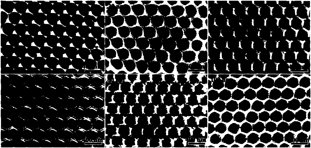
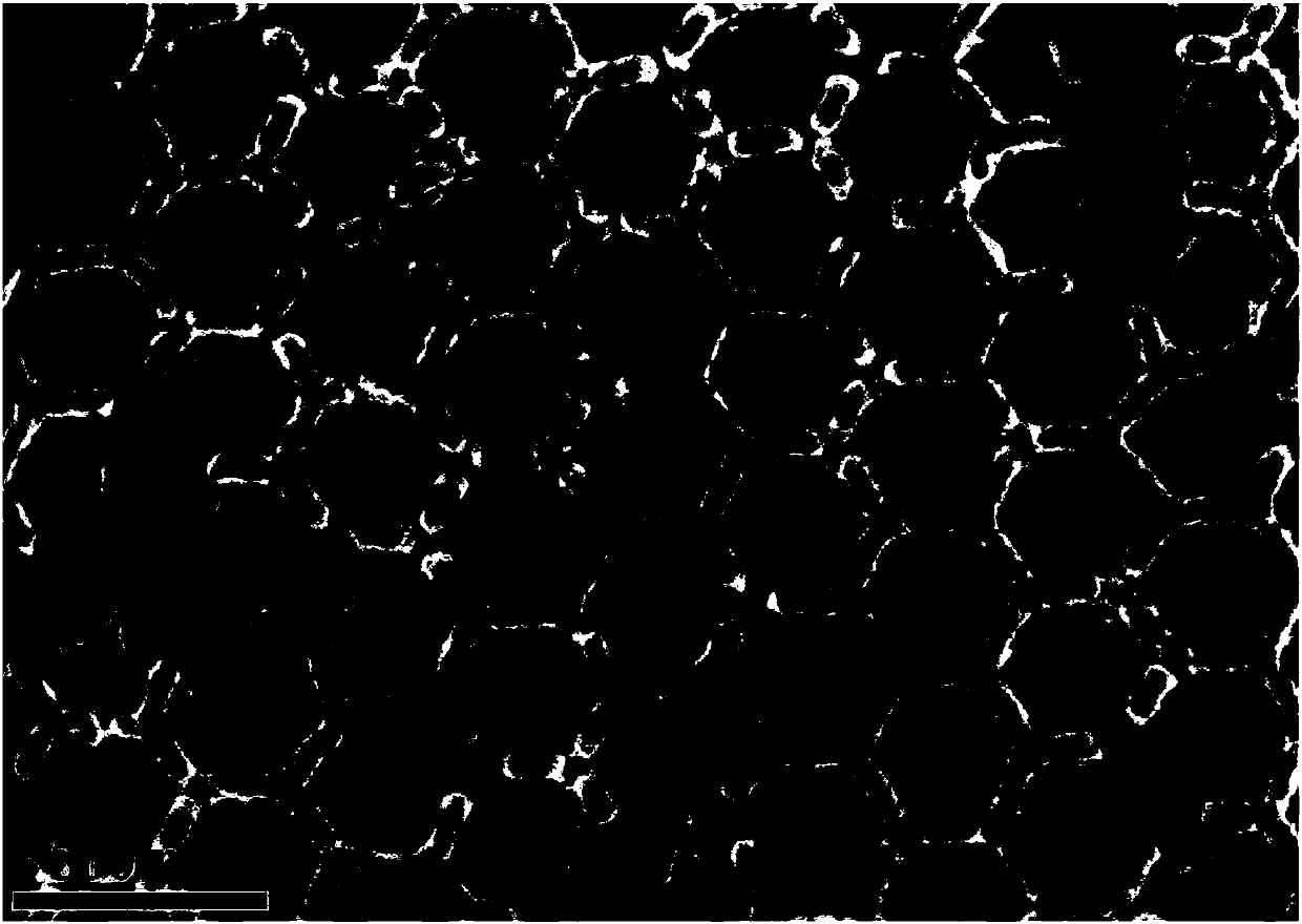
[0027] 1) Spin-coat 50 μL of 5% polystyrene microsphere suspension with a diameter of 400 nm on a 1 cm × 1 cm silicon dioxide substrate by spin coating (the substrate is pre-washed with ethanol, acetone, deionized washed with water for 10 min successively and then used for later use), and dried at 35°C to obtain a multilayer close-packed polystyrene microsphere array as a mask plate. Wherein, the specific conditions of the spin coating method are: the rotating speed is 450r / min, and the time is 30s.

[0028] 2) Dip the multilayer close-packed polystyrene microsphere array into TiO 2 Soak in the precursor solution (tetrabutyl titanate: hydrochloric acid: deionized water = 1:10:10, volume ratio, all chemical solutions used are analytically pure) for 30 minutes and take it out, and then use the spin coating method to remove the excess precursor solution on the surface again. The temperature is 1000r / min, and the time is 30s; then the p...

Embodiment 2

[0036] (1) Prepare SERS substrate 80nm@290nm TiO according to the method in Example 1 2 Invert the opal and cut it into a size of 0.5cm×0.5cm for later use.

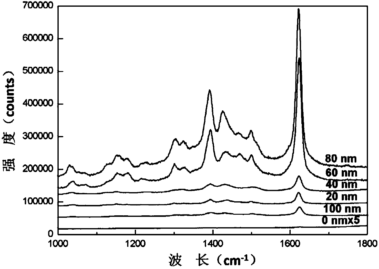
[0037] (2) According to the method in Example 1, the exosomes in the plasma of normal people and prostate cancer patients were extracted, and Raman test was performed.

[0038] Analyze the sample of this embodiment, Figure 5 It is the SERS pattern corresponding to the exosomes of normal people and prostate cancer patients in this example, and the illustration is the corresponding 1087cm -1 Raman peak intensity at different sites (10 to 15 cases of exosomes from normal subjects and patients with benign prostatic hyperplasia). 1087cm -1 The vibration bond corresponding to the phosphate radical, the comparison shows that the degree of phosphorylation of exosomes in patients with prostate cancer is significantly higher than that of normal people, so the degree of phosphorylation of exosome proteins extracted from plasma ...


PUM

PropertyMeasurementUnit
pore sizeaaaaaaaaaa
diameteraaaaaaaaaa
diameteraaaaaaaaaa

Abstract

The invention discloses preparation of an SERS (Surface Enhanced Raman Scattering) substrate and application of the SERS substrate to cancer detection and belongs to the field of nanomaterials. The SERS substrate is TiO2 inverse opal plated with 80nm Au, and the bore diameter of the TiO2 inverse opal is 290nm. In the SERS substrate, the bore diameter of the TiO2 inverse opal is matched with the diameter (about 90nm) of an exosome of a to-be-detected object, and the exosome can make full contact with the SERS substrate; and on the other hand, the TiO2 inverse opal with the bore diameter being 290nm can generate a slow light effect during analyte detection under the excitation of a 633nm laser, and then an SERS signal of the exosome itself can be better obtained. By use of the SERS substrate, the phosphorylation degree of exosome protein extracted from plasma can be quickly analyzed, and then a cancer is diagnosed; consumed time is short, and sensitivity is high; and a new thought is provided for quick diagnosis of a non-invasive in-vitro cancer.

Description

technical field [0001] The invention belongs to the field of nanomaterials, and in particular relates to the preparation of a SERS substrate and its application in analyzing the phosphorylation degree of exosome proteins in blood plasma and then diagnosing cancer. Background technique [0002] Exosomes are small lipid bilayer membrane-wrapped nanovesicles (30-120 nm in diameter) derived from endocytosis. All cells can secrete exosomes, so they are widely present in biological fluids such as saliva, urine, milk and blood. Accumulating evidence shows that exosomes play an important role in tumor biology and metastasis and carry many characteristics of parental cells, such as proteins, DNA and RNA molecules, so their role as cellular messengers has become a cancer and potential biomarkers of disease. Currently, the mainstay of cancer diagnosis is invasive biopsy. The in-depth study of exosomes shows that as a cancer marker, it can be used as a non-invasive means in cancer di...

Claims


Application Information

Patent Timeline
Patent Type & Authority Applications(China)
IPC IPC(8): G01N21/65
CPCG01N21/658
Inventor 肖湘衡董仕练蒋昌忠
Owner WUHAN UNIV
Why Eureka
Try Eureka