Method for extracting vascular matrix component in adipose tissue and application thereof
A technology of adipose tissue and extraction method, which is applied in the direction of medical preparations containing active ingredients, bone/connective tissue cells, drug combinations, etc., and can solve the problems of low stem/progenitor cell yield and limiting the use of SVF
- Summary
- Abstract
- Description
- Claims
- Application Information
AI Technical Summary
Problems solved by technology
Method used
Image
Examples
Embodiment 1
[0038] The preparation method (experimental group) of the SVF of the embodiment of the present invention comprises the following steps:
[0039] Step 1, collecting adipose tissue, adding buffer to wash the adipose tissue. to remove blood cells.
[0040] Use liposuction technology to extract 20 mL of subcutaneous fat tissue from the thigh or abdomen of healthy adult women, cut the fat tissue into pieces mechanically, place in a 50 mL centrifuge tube, add 20 mL of phosphate buffered saline (Phosphate Buffered Saline, PBS), mix well, and let stand After 3 minutes, the lower aqueous phase was discarded by suction, and the washing was repeated 2 to 3 times to remove blood cells.
[0041] Step 2: Add buffer to resuspend adipose tissue, centrifuge and discard the upper layer of fat.
[0042] Add 20mL PBS to resuspend adipose tissue, centrifuge at 4000rpm for 1min, discard the upper layer of fat, and repeat washing 2-3 times to remove the fat.
[0043] Step 3, repeatedly sucking an...
experiment example 1
[0058] Detection of the viability of CD34+ cells in the experimental group and the control group
[0059] Add 5 mL of PBS to resuspend the SVF obtained from the experimental group and the control group, respectively, and sample the CD34+ cell ratio and cell viability by flow cytometry. The test results are shown in Table 1. The proportion of CD34+ cells in the SVF prepared by the experimental group was 32.82%, while the proportion of CD34+ cells in the SVF prepared by the experimental group was only 5.52%. The proportion of CD34+ cells in the experimental group was 6 times that of the control group above. The total viability of cells in the SVF prepared by the experimental group was 93.23%, and the number of viable cells in SVF was 4.53×10 6 / mL. The total viability of cells in the SVF prepared by the control group was 80.57%, and the number of viable cells in SVF was 1.22×10 6 / mL. The adipose tissue and total cell viability obtained per milliliter of the experimental gro...
Embodiment 2
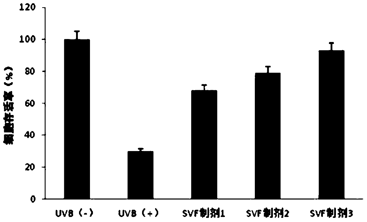
[0065] Preparation of anti-photoaging formulations including SVF
[0066] Using the SVF prepared in Example 1, an anti-photoaging formulation including SVF was formulated.
[0067] SVF preparation 1: Dilute SVF with saline to a concentration of 10 5 / mL, add astragaloside B, retinoic acid, and vitamin C respectively, so that the mass-to-volume ratio is 0.02%, 0.02%, and 0.01%, respectively.
[0068] SVF preparation 2: Dilute SVF with saline to a concentration of 10 6 / mL, add astragaloside B, retinoic acid, and vitamin C respectively, so that the mass-to-volume ratio is 0.03%, 0.03%, and 0.02%, respectively.
[0069] SVF preparation 3: Dilute SVF with saline to a concentration of 10 7 / mL, add astragaloside B, retinoic acid, and vitamin C respectively, so that the mass-to-volume ratio is 0.05%, 0.05%, and 0.03%, respectively.
PUM
 Abstract
Description
Claims
Application Information
 - R&D
- Intellectual Property
- Life Sciences
- Materials
- Tech Scout
- Unparalleled Data Quality
- Higher Quality Content
- 60% Fewer Hallucinations
Browse by: Latest US Patents, China's latest patents, Technical Efficacy Thesaurus, Application Domain, Technology Topic, Popular Technical Reports.
© 2025 PatSnap. All rights reserved.Legal|Privacy policy|Modern Slavery Act Transparency Statement|Sitemap|About US| Contact US: help@patsnap.com


