

Separation method of human placenta sub-totipotent stem cells
A sub-totipotent stem cell and separation method technology, applied in the direction of cell dissociation methods, embryonic cells, animal cells, etc., can solve the problems of unclean collection, affecting the effect of enzymatic hydrolysis, and many miscellaneous cells, so as to reduce the separation time and improve the separation Efficiency, the effect of ensuring the effect of enzymatic hydrolysis
- Summary
- Abstract
- Description
- Claims
- Application Information
AI Technical Summary
Problems solved by technology
Method used
Image

Examples
Embodiment 1
[0050] The junction of the umbilical cord and the placenta is used as a dot, and the line connecting the apex and the edge of the placenta is used as the axis circle, and the amniotic membrane is removed to obtain a circular amniotic membrane sample with a width of 3-5cm.
[0051] Rinse the removed circular placental amniotic membrane three times with phosphate buffer solution with a pH value of 7.3, then put it into a kidney-shaped dish, and use tweezers and a cell scraper to clean the clots and gelatinous substances on the membrane to obtain amniotic membrane Sample, to prevent affecting the digestion efficiency of the enzyme; the clot on the placenta is a black-red block that adheres to the membrane, and the jelly is a transparent viscous substance that is attached to the surface of the amniotic membrane. It is used in a kidney-shaped disc. Use tweezers to remove large pieces of coagulated blood, and then use a cell scraper to gently and slowly scrape off the remaining coagu...
Embodiment 2
[0058] The amniotic membrane was randomly stripped from the placenta, washed repeatedly with D-Hank’s balanced salt solution with pH=7.3 to remove residual blood, and the amniotic membrane was cut into 5×5cm pieces 2 Small piece;
[0059] Drop 1mL of D-Hank's balanced salt solution into a 6-cm petri dish, wet the bottom of the petri dish, place the epithelial layer of the amniotic membrane down in the petri dish, use slides to separate the gelatinous substance of the mesenchymal layer scrape off;
[0060] Put the scraped amniotic membrane into 10 mL of D-Hank's balanced salt solution containing 10 (w / v)% N-acetyl-L-cysteine, and incubate at room temperature for 15 minutes; remove the residual mucus on the amniotic membrane for easy digestion completely;
[0061] Add 20mL of protease solution to the amnion, digest it in an air bath at 37°C for 30min, collect the digestion solution and add 5mL of fetal bovine serum; wherein the protease solution is 0.15 (w / v)% collagenase II-E...
Embodiment 3
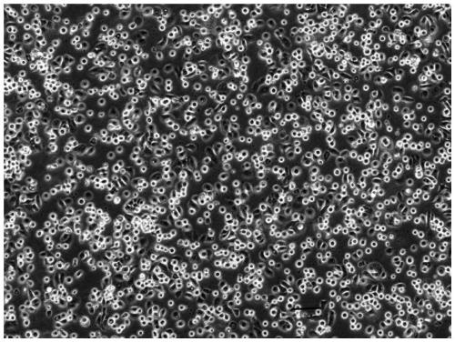
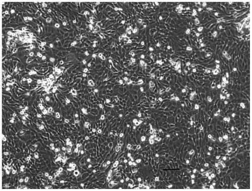
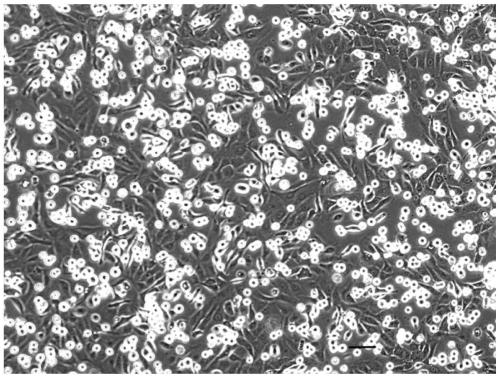
[0069] The cell suspensions of the three control groups were all grown in vitro, and the cell growth was observed with a microscope after three days and five days. The growth of the first control group was as follows: Figure 1-Figure 4 shown;
[0070] Because the shape of placental subtotipotent stem cells is round, Figure 1-3 The shape of the cells is basically round, and the Figure 4 There are spindle cells in it, it can be seen that the cells separated by the patent method are more pure than the human placental subtotipotent stem cells separated by the traditional method.
PUM
| Property | Measurement | Unit |
|---|---|---|
| width | aaaaa | aaaaa |
Abstract
Description
Claims
Application Information