Applications of uterine cavity fluid-derived exosome in preparation of drugs and adjuvant therapeutic agents for treating infertility related diseases

A technology for infertility and adjuvant treatment, applied in the direction of diseases, drug combinations, microorganisms, etc., can solve the problems of unspecified use of exosomes, application research of exosomes without source, etc., to achieve good therapeutic effect, The effect of simple preparation process and broad clinical application prospects

Active Publication Date: 2020-04-03
PHARCHOICE THERAPEUTICS INC
View PDF2 Cites 4 Cited by
  • Summary
  • Abstract
  • Description
  • Claims
  • Application Information

AI Technical Summary

Problems solved by technology

Patent application 201811552777.3 discloses a method for extracting uterine cavity fluid exosomes from living pigs, but fails to clarify the use of this type of exosomes, and there is no application research on exosomes from this source in the prior art

Method used


Image

  • Applications of uterine cavity fluid-derived exosome in preparation of drugs and adjuvant therapeutic agents for treating infertility related diseases
  • Applications of uterine cavity fluid-derived exosome in preparation of drugs and adjuvant therapeutic agents for treating infertility related diseases
  • Applications of uterine cavity fluid-derived exosome in preparation of drugs and adjuvant therapeutic agents for treating infertility related diseases

Examples


Embodiment 1

[0029] Example 1 Preparation of uterine cavity fluid exosomes

[0030] The uterine cavity fluid from healthy volunteers was filtered with a 0.45 μm filter membrane, centrifuged at 4°C, 1000g for 10min, and the supernatant was collected; the collected supernatant was centrifuged at 4°C, 2000g for 20min, and the supernatant was collected; the collected supernatant was 4°C , centrifuge at 10000g for 30min, collect the supernatant; centrifuge the collected supernatant at 110000g for 100min, discard the supernatant, and resuspend the pellet with phosphate buffer; Filter with a 0.45 μm filter membrane to obtain exosomes. The total protein content of exosomes was detected by Bradford method (Bio-RadProteinAssayReagent). Exosomes were stored frozen at -80°C. Exosomes were extracted from the same healthy volunteer serum in the same way as control exosomes.

Embodiment 2

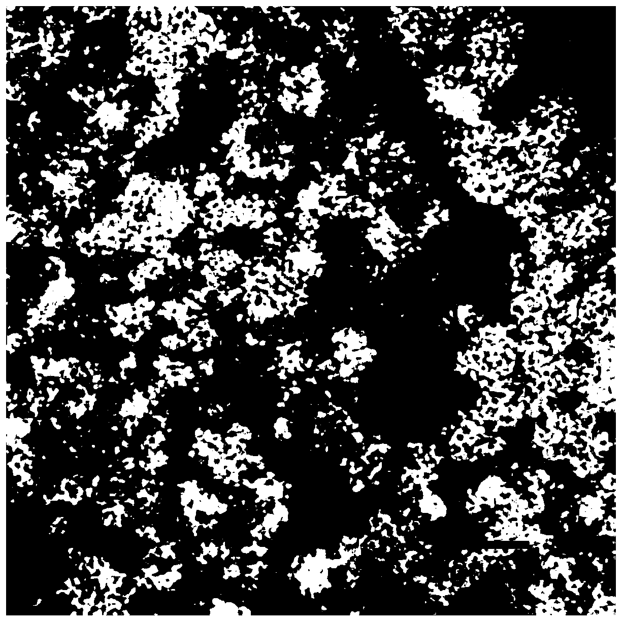
[0031] Example 2 Exosome electron microscope detection

[0032]Exosome electron microscope detection was carried out according to the following steps: thaw the exosomes of uterine cavity fluid obtained in Example 1 and resuspend the sample with buffer solution; drop the sample on a copper grid with a support membrane; mix the dye solution with the sample Mix evenly to obtain a mixed solution. After the suspension on the copper grid is slightly dried, drop the mixed solution on the copper grid, negatively stain for 2 minutes, and then air-dry naturally; observe under a transmission electron microscope. Double-membrane vesicles consistent with the 30-150nm morphological characteristics of exosomes were observed, such as figure 1 shown.

Embodiment 3

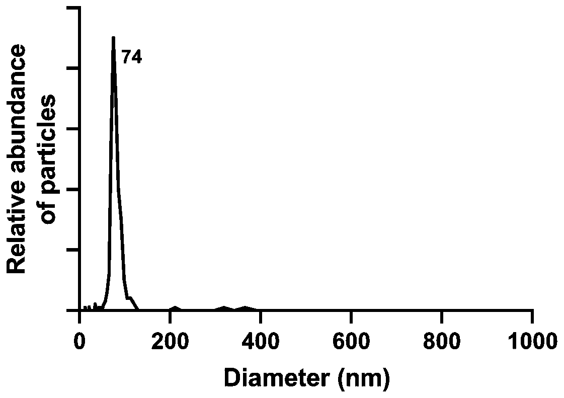
[0033] Example 3 Exosome particle size analysis

[0034] Utilize NanoFCM Flow NanoAnalyzer (nano particle size analyzer) to carry out particle size detection method to the uterine cavity fluid exosome obtained in embodiment 1, specifically comprise the following steps:

[0035] Particle size detection: Particle size standard, blank control and sample to be tested (uterine cavity fluid) all need to be sampled under the same detection conditions (laser power and scattering channel attenuation coefficient are exactly the same); all samples are under lower injection pressure (sampling is not greater than 1.0 kPa) detection; detect the particle size standard under suitable conditions (the scattered light signal of the smallest silicon sphere is completely separated from the background and the largest silicon sphere signal is not saturated, and the detector saturation value is 3.6k ); detect the buffer used to resuspend the sample to be tested, used to subtract background particles,...


PUM

PropertyMeasurementUnit
diameteraaaaaaaaaa
diameteraaaaaaaaaa
particle diameteraaaaaaaaaa

Abstract

The invention relates to the technical field of medicines, and provides applications of uterine cavity fluid-derived exosome in preparation of drugs and adjuvant therapeutic agents for treating infertility related diseases. Through the verification of experiments, exosome realizes the treatment or relieving of endometrial growth disorders by promoting the increasing of the thickness of endometrium, enhancing the activity of endometrial cells, reducing the damages to intimal cells, promoting VEGF expression, maintaining the dryness of endometrial stromal cells and stimulating proliferation; andthrough the realizing of the treating or relieving of maternal-fetal immune tolerance disorder-related diseases by achieving immune tolerance effects, treating spontaneous abortion and enhancing T helper lymphocyte levels, a novel theoretical basis can be provided for infertility treatment. In addition, for both fertilized eggs being fertilized in vivo or fertilized in vitro, the exosome can effectively enhance developmental rates in multiples; and implantation rates and farrowing rates of in vitro fertilization transplantation can be increased as well, so that positive auxiliary effects on infertility treatment can be achieved.

Description

technical field [0001] The invention belongs to the field of biomedicine, and relates to the use of exosomes derived from uterine cavity fluid in the preparation of drugs and auxiliary therapeutic agents for infertility-related diseases, a preparation method of the exosomes, and a pharmaceutical composition containing the exosomes . Background technique [0002] The endometrial injury is mainly the injury of the endometrial basal layer, which is mainly related to curettage during pregnancy. Compared with the normal endometrium, the endometrial basal layer during pregnancy is loose and more susceptible to damage. Based on my country's national conditions and the increasing rate of induced abortion, the occurrence of endometrial damage cannot be ignored. The endometrial basal layer is damaged, which may lead to damage or loss of endometrial stem cells; at the same time, local infection and aseptic inflammation of the injured endometrium can destroy the niche microenvironment ...

Claims


Application Information

Patent Timeline
Patent Type & Authority Applications(China)
IPC IPC(8): A61K35/48A61P15/08A61P15/06C12N5/071
CPCA61K35/48A61P15/06A61P15/08C12N5/0682C12N2509/00
Inventor 胡适丁敏
Owner PHARCHOICE THERAPEUTICS INC
Why Eureka
Try Eureka