Method, primers and kit for detecting PD-L1 gene expression quantity through real-time fluorescent quantitative PCR

A PD-L1 and gene technology, applied in the field of real-time fluorescence quantitative PCR detection of PD-L1 gene expression, primers and kits, can solve the problems of incomplete and accurate reflection of the expression of the gene to be tested, unstable expression, etc. Achieve the effect of eliminating instability, avoiding subjectivity, and improving sensitivity

Pending Publication Date: 2020-04-17
江苏普瑞悉恩生物科技有限公司 +1
View PDF5 Cites 1 Cited by
  • Summary
  • Abstract
  • Description
  • Claims
  • Application Information

AI Technical Summary

Problems solved by technology

[0007] Traditional RNA expression detection methods usually use GAPDH, beta-actin, tubulin and other mRNAs as references. However, due to the different expression levels of reference genes in different tissue p...

Method used


Image

  • Method, primers and kit for detecting PD-L1 gene expression quantity through real-time fluorescent quantitative PCR
  • Method, primers and kit for detecting PD-L1 gene expression quantity through real-time fluorescent quantitative PCR
  • Method, primers and kit for detecting PD-L1 gene expression quantity through real-time fluorescent quantitative PCR

Examples


Embodiment 1

[0076] Example 1 Extraction of RNA / DNA from MDA-MB-231 cells, K562 cells and tumor tissue samples treated with interferon-γ

[0077] Using the AllPrep DNA / RNAMini Kit, the DNA / RNA extraction kit can extract up to 1*10 7 Cells or 30mg tissue.

[0078] Cells: cell number ≤1×10 7 , add 600μL Buffer RLTPlus; tissue: tissue ≤ 30mg, add 600μL Buffer RLTPlus. The lysate was then centrifuged at top speed for 3 minutes and the supernatant was carefully removed using a pipette.

[0079] Transfer to an Allprep DNA adsorption column (placed in a 2mL collection tube), and centrifuge at 10,000rpm for 30 seconds.

[0080] RNA extraction: add an equal volume of 70% ethanol to the centrifuged liquid from the above-mentioned Allprep DNA adsorption column, and mix well.

[0081] Pipette 700uL into the RNeasy adsorption column (placed in a 2mL collection tube), centrifuge at 10,000rpm for 15 seconds, and discard the waste liquid in the collection tube. Repeat until all RNA is collected.

[...

Embodiment 2

[0091] Embodiment 2 carries out reverse transcription reaction to extraction sample RNA / DNA

[0092] Configure the following mixture in RNA-free Microtube:

[0093] Table 1, cDNA reverse transcription system 1

[0094]

[0095] Place in a water bath at 65°C for 5 minutes, and cool on ice.

[0096] Prepare the following reaction solution in the above tube to a total volume of 20uL, and carry out the reverse transcription reaction.

[0097] Table 2, cDNA reverse transcription system 2

[0098]

[0099] Mix gently and place in a water bath at 45°C for 60 minutes.

[0100] Place in a water bath at 70°C for 15 minutes and cool on ice.

[0101] Obtain a cDNA / DNA solution sample for the next step of fluorescent quantitative PCR reaction.

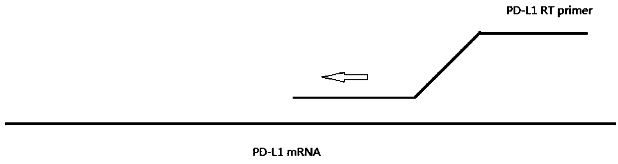
[0102] figure 1 is a schematic diagram of the reverse transcription reaction method, represented by figure 1 It can be seen that RNA is used as a template, PD-L1RT is used as a specific primer, and cDNA is obtained by reaction under th...

Embodiment 3

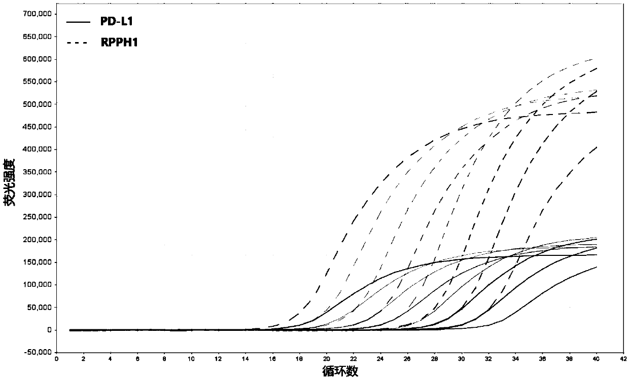
[0103] Example 3 Carry out dual-channel fluorescent quantitative PCR reaction and result calculation for cDNA / DNA samples

[0104] Configure the mixed solution according to the following reaction system (25uL / tube)

[0105] Table 3. Real-time quantitative PCR reaction system

[0106]

[0107] Fluorescent quantitative PCR reaction:

[0108] Table 4. Real-time quantitative PCR reaction conditions

[0109]

[0110]

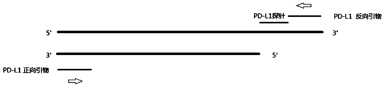
[0111] figure 2 It is the amplification method of PD-L1 cDNA fluorescent quantitative PCR, which is composed of figure 2 It can be seen that during PCR amplification, the 5'-3' exonuclease activity of Taq enzyme will digest and degrade the probe, so that the fluorescent emitting group and the fluorescent quenching group are separated, so that the fluorescent monitoring system can receive the fluorescent signal, that is Every time a DNA strand is amplified, a fluorescent molecule is formed, which realizes the complete synchronization of the accumulation...


PUM


Abstract

The invention discloses a method, primers and a kit for detecting PD-L1 gene expression quantity through real-time fluorescent quantitative PCR. The method comprises the following steps: (1) synchronously extracting DNA and mRNA in a sample; (2) synthesizing a first chain cDNA by taking the to-be-detected mRNA as a template through a special reverse transcription primer; and (3) carrying out dual-channel real-time fluorescent quantitative PCR amplification by respectively adopting a primer corresponding to the specific sequence on the reverse transcription primer and a specific primer corresponding to a reference gene and taking a reverse transcription product cDNA and reference gene DNA as templates while amplifying a target gene cDNA and a reference gene, and calculating the copy numberratio of the target gene to the reference gene in the sample according to the respective standard curves by taking a plasmid constructed by linking the reference gene and the target gene sequence as astandard substance to obtain the expression level of the target gene, so that various experimental operation errors are synchronously corrected, and the reliability and the repeatability of the result are ensured.

Description

technical field [0001] The invention relates to a method for detecting the expression level of PD-L1 gene. Background technique [0002] Programmed cell death ligand 1 (processed death-1ligand 1, PD-L1), also known as B7-H1, CD274, is an inhibitory co-stimulatory molecule encoded by the CD274 gene. PD-L1 is a type I transmembrane glycoprotein with 290 amino acids, including an extracellular region, a hydrophobic transmembrane region and a tail cytoplasmic region. PD-L1 protein is widely expressed in lymphocytes, such as activated T and B cells, macrophages, dendritic cells, thymic endothelial cells and other cells, as well as in the heart and placenta, as well as in various tumor cells and tumor microenvironments. Highly expressed in immune cells in lung cancer, esophageal cancer, breast cancer, lymphoma, etc., the PD-L1 described in the literature: Dong, H. et al. (2002) NatMed 8, 793-800, hompson, R.H. et al. ( 2006) Cancer Res 66, 3381-5. and Pardoll, D.M. (2012) Nat Re...

Claims


Application Information

Patent Timeline
IPC IPC(8): C12Q1/6886C12N15/11C12Q1/6851
CPCC12Q1/6886C12Q1/6851C12Q2600/166C12Q2600/136C12Q2531/113C12Q2561/113C12Q2563/107
Inventor 唐向荣唐志君张冀申
Owner 江苏普瑞悉恩生物科技有限公司
Why Eureka
Try Eureka