Fusion protein of monomeric streptavidin and Gaussian luciferase and its application

A technology of streptavidin and fusion protein, applied in the field of fusion protein of monomer streptavidin and Gaussian luciferase, can solve the problems of substrate aggregation, time-consuming, cumbersome process, etc. The effect of simple production and production process and broad application prospects

Active Publication Date: 2022-02-22
青岛华大智造科技有限责任公司
View PDF1 Cites 0 Cited by
  • Summary
  • Abstract
  • Description
  • Claims
  • Application Information

AI Technical Summary

Problems solved by technology

This method is cumbersome and time-consuming
And if SA and anti-SA antibodies are used, it may also cause substrate aggregation

Method used


Image

  • Fusion protein of monomeric streptavidin and Gaussian luciferase and its application
  • Fusion protein of monomeric streptavidin and Gaussian luciferase and its application
  • Fusion protein of monomeric streptavidin and Gaussian luciferase and its application

Examples


Embodiment 1

[0099] Embodiment 1: Construction of fusion expression vector

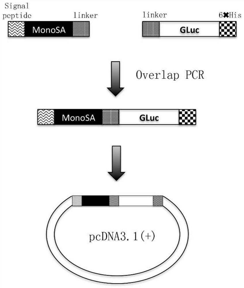
[0100] Such as figure 1 As shown, the MonoSA gene and the Gaussian luciferase gene were used as templates to design and synthesize primers for PCR amplification to obtain the MonoSA and Gaussian luciferase gene amplification products respectively. Based on this PCR product, a second round of bridging PCR was performed to obtain the amplified product of the fusion gene of MonoSA and Gaussian luciferase (sequence such as SEQ ID NO: 13). The PCR amplified product was detected by electrophoresis on 1% agarose gel, and the product was recovered with a gel recovery kit. The recovered product and the vector plasmid pcDNA3.1(+) were subjected to a double enzyme digestion reaction with EcoR I and Hind III respectively. After the reaction was completed, electrophoresis was performed on a 1% agarose gel for detection, and the product was recovered with a gel recovery kit . The products recovered from PCR digestion and the...

Embodiment 2

[0102] Example 2: Transfection, expression and purification of fusion expression vector

[0103] Mix 25 μg of the constructed pcDNA3.1-MonoSA-Gluc fusion expression plasmid with 1 ml of cell culture medium evenly. Another 920 μL cell culture medium was mixed with 80 μL lipo2000 transfection reagent (invitrogen). Mix the diluted expression vector and the diluted transfection reagent, and let stand at room temperature for 20 minutes. Prepare 25ml concentration of 2×10 6 / ml cells, add the mixed pcDNA3.1-MonoSA-Gluc fusion expression plasmid and transfection reagent mixture into CHO cells, and shake gently. 37°C, 8% CO 2 conditions for 3 days.

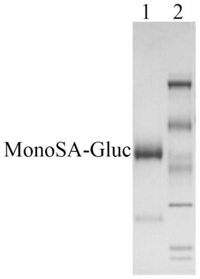
[0104] Collect the cell culture fluid, centrifuge at 6000rpm for 10min, and collect the supernatant. After the supernatant was filtered, it was passed on to a nickel column, and 10 ml of PBS was equilibrated. Then wash 10 ml with buffer (20mM Tris, pH 8.0, 150mM NaCl, 20mM imidazole) to remove foreign proteins, and finally elute wit...

Embodiment 3

[0106] Example 3: Fusion protein luciferase bioluminescent detection

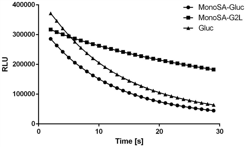
[0107] MonoSA-Gluc fusion protein and MonoSA-G2L fusion protein were diluted to 3nM with substrate diluent (50mM Tris, pH 8.0, 100mM NaCl), respectively, and 10 μL was added to a 96-well microtiter plate. Then add 90 μL of the substrate coelenterazine diluted to 10 μM with the same solution, and read the luminous intensity with the self-luminescence module of the microplate reader, and the results are as follows image 3 As shown, the luminescence intensity of wild-type MonoSA-Gluc fusion protein is equivalent to that of Gluc protein at the same concentration. The luminescence intensity of the mutant fusion protein MonoSA-G2L fusion protein was comparable to that of Gluc, but it obviously showed continuous luminescence characteristics, and the luminescence did not weaken significantly after 30s.


PUM


Abstract

A fusion protein of monomeric streptavidin and Gaussian luciferase and its application, the fusion protein comprises monomeric streptavidin full-length protein and Gaussian luciferase full-length protein or continuous light-emitting mutant, single The streptavidin full-length protein is connected to the Gaussian luciferase full-length protein or the persistent light-emitting mutant through a flexible peptide. The fusion protein of the invention can recognize the biotin molecule conveniently and specifically, and has the function of catalyzing the oxidation and luminescence of the substrate, which is convenient for detection. Compared with the combination of SA and anti-SA antibody to detect biotin, this fusion protein is not only convenient and fast, but also does not cause the aggregation of the substrate, and only needs one expression and purification to obtain the fusion protein. The production process is simple, which is beneficial to mass production. The fusion protein has broad application prospects in the fields of DNA hybridization, immune detection, biochemical diagnosis and the like.

Description

technical field [0001] The invention relates to the field of genetic engineering, in particular to a fusion protein of monomeric streptavidin and Gaussian luciferase and its application. Background technique [0002] Streptavidin (streptavidin, SA) was discovered by Chaiet in 1963 and is a product secreted by Streptomycesavidinii bacteria during the culture process. There are 159 amino acid residues in a SA peptide chain, and the molecular weight is 16.5kDa. The complete SA protein consists of 4 SA polypeptide chains, forming a tetrameric structure. Each polypeptide chain can specifically bind one biotin molecule, therefore, one molecule of SA can bind four molecules of biotin. The interaction between the two exists in the form of non-covalent binding, and the affinity reaches 10 -15 / M, which is several orders of magnitude higher than the binding constant of antigen and antibody, is the most stable protein-small molecule interaction existing in nature. The strong bindin...

Claims


Application Information

Patent Timeline
Patent Type & Authority Patents(China)
IPC IPC(8): C07K19/00C12N15/62C12N15/85C12N5/10G01N33/82
CPCC07K14/36C12N15/85G01N33/82C07K2319/61C07K2319/21C07K2319/02
Inventor 李静王佑富郑越董宇亮章文蔚徐崇钧
Owner 青岛华大智造科技有限责任公司
Why Eureka
Try Eureka