Kit and method for detecting programmed cell death 1 ligand 1 (PD-L1) mRNA on tissue

A kit and tissue technology, applied in the field of kits for detecting PD-L1mRNA on tissue, can solve problems such as false negatives, affecting probe penetration, unreasonable probe design, etc.

Active Publication Date: 2020-07-07
ZHENGZHOU KODIA BIOTECHNOLOGY CO LTD
View PDF4 Cites 0 Cited by
  • Summary
  • Abstract
  • Description
  • Claims
  • Application Information

AI Technical Summary

Problems solved by technology

However, the accuracy of FISH detection is not high at present, and there are problems of false positives and false negatives.
[0005] Reasons for false positives in FISH detection include (a) unreasonable probe design; (2) interference from microbial autofluorescence; (c) natural fluorescent biological or chemical substances in environmental samples (such as activated sludge or drinking water, etc.) Interference of residues; (d) Effects of

Method used


Image

  • Kit and method for detecting programmed cell death 1 ligand 1 (PD-L1) mRNA on tissue
  • Kit and method for detecting programmed cell death 1 ligand 1 (PD-L1) mRNA on tissue
  • Kit and method for detecting programmed cell death 1 ligand 1 (PD-L1) mRNA on tissue

Examples


Embodiment 1

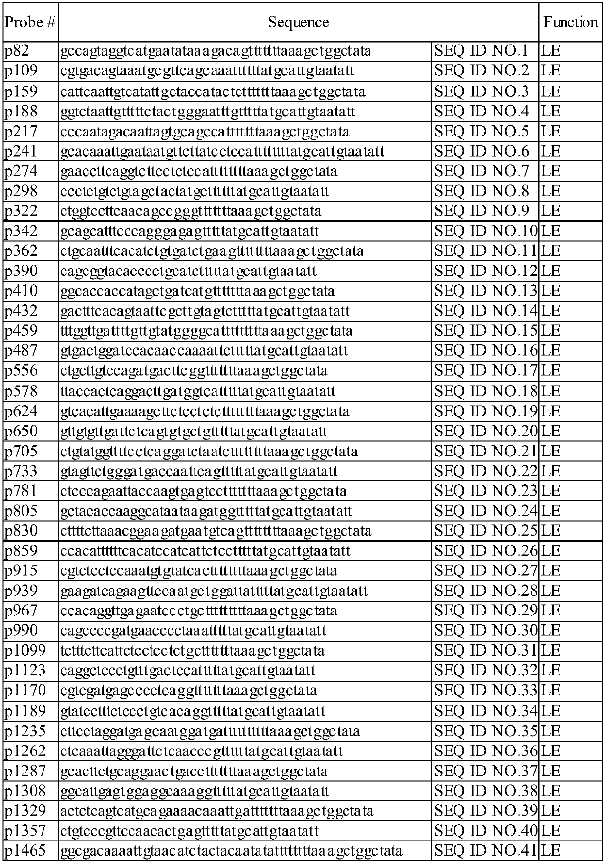
[0028] Example 1. Probe design

[0029] By comparing the PD-L1 mRNA sequence, design and synthesize multiple PD-L1 specific probes, as shown in Table 1. The probe design follows the principle of pairwise pairing and cooperative capture. All in situ hybridized cDNA probes do not exist in isolation, and need to be paired with paired probes to combine with the branched DNA amplification tree structure.

[0030] Table 1 PD-L1 specific probes

[0031]

[0032]

[0033]

Embodiment 2

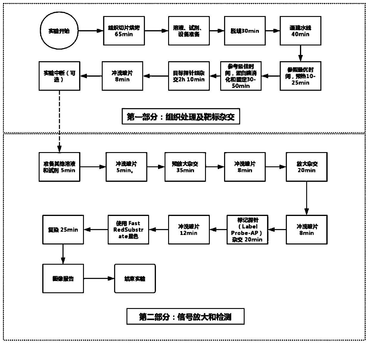
[0034] Embodiment 2. Detection method

[0035] 1. Pretreatment of tissue and hybridization of target

[0036] 1. Bake tissue slices at 60℃ for 1h.

[0037] 2. Prepare the following liquid in the gap between the baked slices:

[0038] (1) Prepare 2L 1×PBS: 1.8L ddH 2 O+0.2L 10×PBS;

[0039] (2) Prepare 200ml 10% NBF: 178ml 1×PBS+22ml 37% formaldehyde in the fume hood;

[0040] (3) Prepare 3L 1× washing buffer: 2.94L ddH 2 O+60ml 50× washing buffer, this washing buffer can be kept at room temperature

[0041] Deposit for 1 month;

[0042] (4) Prepare 500ml 1×pretreatment solution: 495ml ddH 2 O+5ml 100×pretreatment liquid;

[0043] (5) Prepare the following reagents:

[0044] 400ml xylene, 400ml 100% ethanol

[0045] (6) Preheat 1×PBS and probe dilution to 40°C. Preheat the substrate enhancement solution and the substrate dilution solution to room temperature.

[0046] 3. 30min dewaxing:

[0047] Do the following in the fume hood:

[0048] (1) Pour each 200ml xylene into two washing liquid tanks....

Embodiment 3

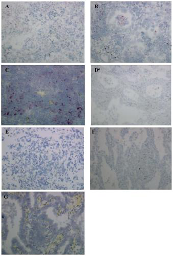
[0112] The 7 cases of lung adenocarcinoma tissues were tested according to the above experimental procedures, and Dako’s PD-L1 antibody 22C3 antibody was used for testing. The results are shown in figure 2 . The statistical results are shown in Table 3.

[0113] Table 3 Test results of 7 cases of lung adenocarcinoma

[0114] Lung adenocarcinoma tissue sample PD-L1 mRNA test results PD-L1 223C antibody test results A2+++ B2++ C3+++ D-- E-- F-- G-+

[0115] The results showed that 3 out of 7 lung adenocarcinoma tissues showed high expression of PD-L1mRNA ( figure 2 ), the conformity of the expression level is equivalent to the detection result of the protein level.


PUM


Abstract

This application belongs to the technical field of biology, and discloses a kit and method for detecting programmed cell death 1 ligand 1 (PD-L1) mRNA on tissue. The kit disclosed by the invention comprises probe sets shown by SEQ ID NO. 1-91. The kit has simple composition, remedies the current situation that immunohistochemical effective antibodies are lacking, eliminates the shortcomings of a fluorescence in situ hybridization (FISH) technology, and greatly expands the field of detection of molecular pathology.

Description

Technical field [0001] The invention belongs to the field of biotechnology, and specifically relates to a kit and method for detecting PD-L1 mRNA on tissues. Background technique [0002] Programmed cell death 1 ligand 1 (PD-L1) is also called cluster of differentiation 274 (CD274) or B7 homolog 1 (B7-H1), It is a protein in the human body, encoded by the CD274 gene. .PD-L1 is the first type transmembrane protein with a size of 40kDa, which is believed to be related to the suppression of the immune system in certain special situations (such as pregnancy, tissue transplantation, autoimmune diseases, and certain diseases such as hepatitis) . Under normal circumstances, the immune system will react to foreign antigens gathered in the lymph nodes or spleen to promote antigen-specific cytotoxic T cells (CD8+Tcell proliferation). The programmed cell death receptor-1 (PD-1) combined with programmed cell death-ligand 1 (PD-L1) can transmit inhibitory signals, reduce the proliferation ...

Claims


Application Information

Patent Timeline
IPC IPC(8): C12Q1/6841C12N15/11
CPCC12Q1/6841C12Q2521/525C12Q2525/207C12Q2521/537Y02A50/30
Inventor 黄瑾张鹭鹭李先坤刘丽杰田小强徐文茹王艺杰孟歌
Owner ZHENGZHOU KODIA BIOTECHNOLOGY CO LTD
Why Eureka
Try Eureka