Eureka AIR delivers breakthrough ideas for toughest innovation challenges, trusted by R&D personnel around the world.

Multilayer microneedle array and preparation method thereof

A microneedle array and single-layer technology, applied in the field of biomedical materials, can solve the problems of great influence on adhesion ability, drop of needle tip, complicated preparation method, etc., and achieve good tissue adhesion, strong repeatability, and simple method Effect

Active Publication Date: 2020-07-31
NANJING DRUM TOWER HOSPITAL
View PDF8 Cites 12 Cited by
  • Summary
  • Abstract
  • Description
  • Claims
  • Application Information

AI Technical Summary

Problems solved by technology

One is to use adhesive materials (such as dopamine gel, polyacrylamide gel, etc.) as the substrate of microneedles, which can stick to the tissue surface through molecular interactions such as hydrogen bonds; The adhesion ability of microneedles is greatly affected by the surrounding environment of the tissue
The second is to use an expandable material as the tip of the microneedle. After contacting the tissue fluid, the needle tip can expand rapidly and mechanically interlock with the tissue to achieve tissue adhesion; however, due to the different materials of the substrate and the needle tip, this type of The tip of the microneedle easily falls off the substrate
The third strategy is to design microneedle tips with special microstructures (for example: barbed structure, arrowhead structure), microneedles can be firmly attached to the tissue with the help of these special physical structures, the disadvantage of this type of microneedle is the preparation method complex

Method used


Image

  • Multilayer microneedle array and preparation method thereof
  • Multilayer microneedle array and preparation method thereof
  • Multilayer microneedle array and preparation method thereof

Examples


Embodiment 1

[0030] Preparation of monolayer microneedle arrays:

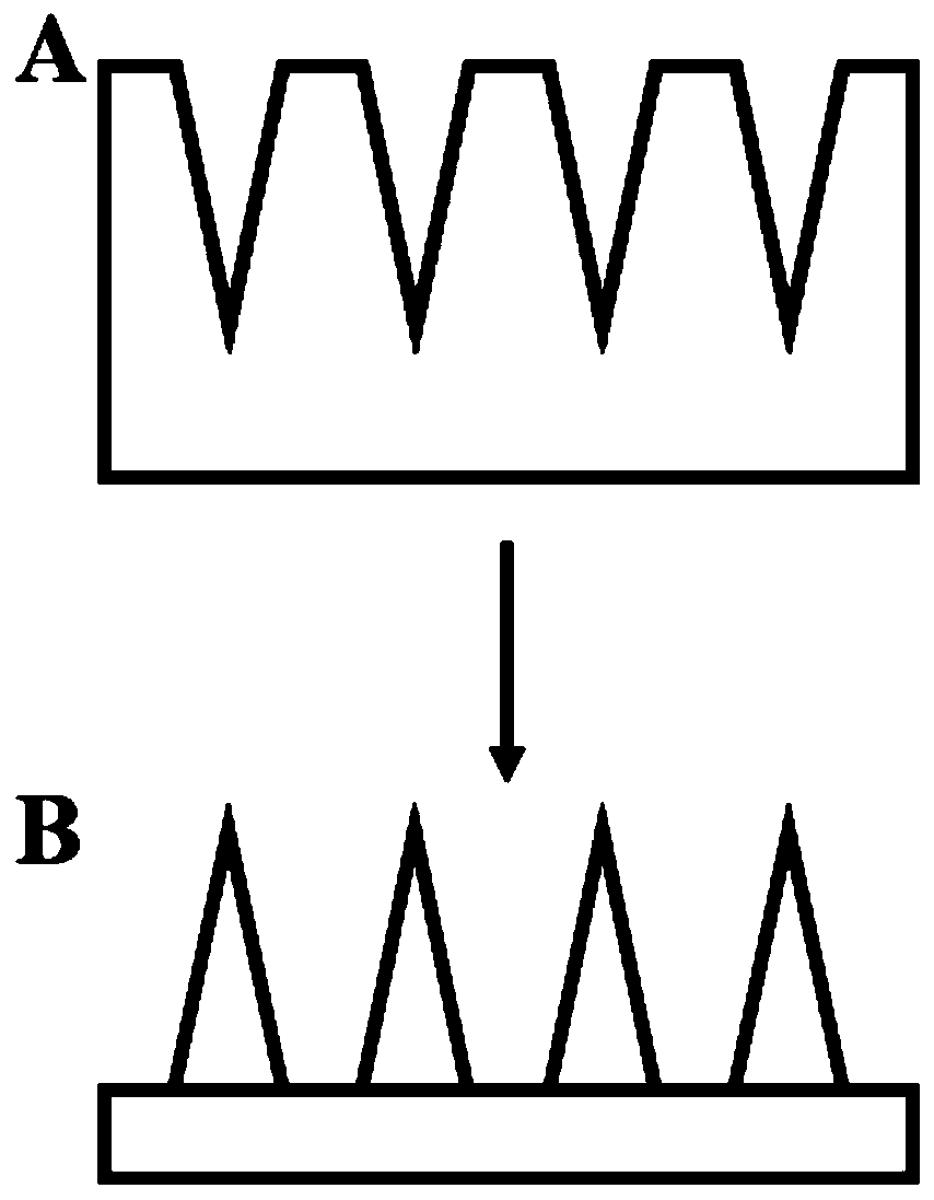
[0031] An aqueous solution containing 50% by volume of polyethylene glycol diacrylate and 1% by volume of 2-hydroxy-2-methylpropiophenone was prepared as a raw material solution. Use a pipette gun to drop 500 microliters of the raw material solution on the polydimethylsiloxane template, and vacuum it for 2 minutes, so that the raw material solution is fully filled in the conical holes of the template (the size of the holes is 400 microns apart, and the depth 500 microns, 350 microns in diameter). After 30 seconds of UV treatment, the raw material solution is cured and then carefully peeled off from the polydimethylsiloxane template, and finally a single-layer microneedle array can be obtained, as figure 1 shown.

Embodiment 2

[0033] Preparation of double-layer microneedle arrays:

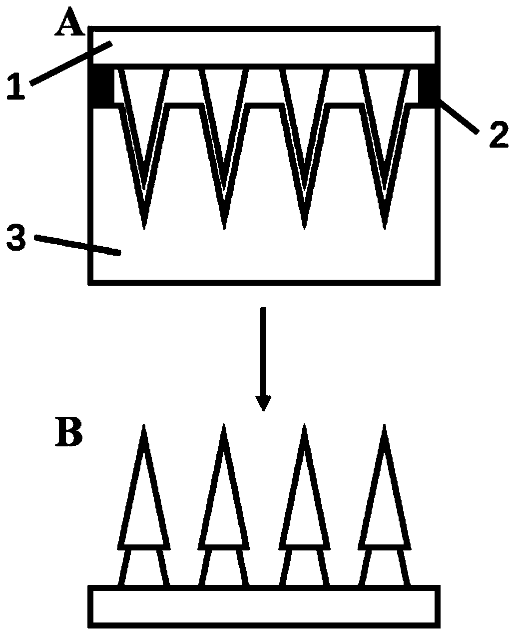
[0034] An aqueous solution containing 50% by volume of polyethylene glycol diacrylate and 1% by volume of 2-hydroxy-2-methylpropiophenone was prepared as a raw material solution. Use a pipette gun to drop 500 microliters of the raw material solution on the polydimethylsiloxane template, and vacuum it for 2 minutes, so that the raw material solution is fully filled in the conical holes of the template (the size of the holes is 400 microns apart, and the depth 500 microns, 350 microns in diameter). After 30 seconds of UV treatment, the raw material solution was cured and then carefully peeled off from the polydimethylsiloxane template to obtain a single-layer microneedle array.

[0035] 500 microliters of the raw material solution was added dropwise on the polydimethylsiloxane template with a pipette gun again, and treated in vacuum for 2 minutes, so that the raw material solution was fully filled in the conical holes of ...

Embodiment 3

[0037] Preparation of three-layer microneedle array:

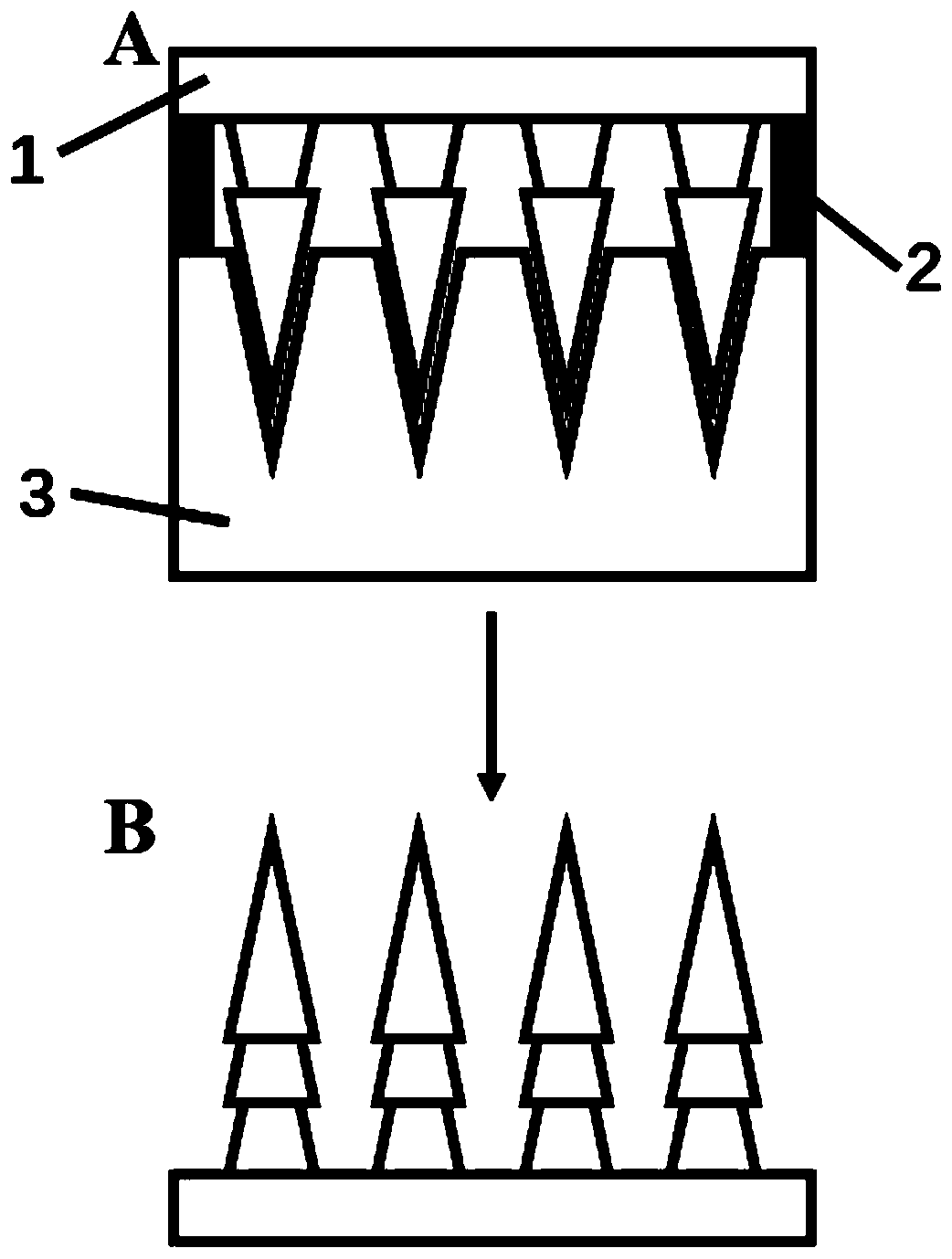
[0038] An aqueous solution containing 30% by volume of methacrylate gelatin and 1% by volume of 2-hydroxy-2-methylpropiophenone was prepared as a raw material solution. Use a pipette gun to drop 500 microliters of the raw material solution on the polydimethylsiloxane template, and vacuum it for 2 minutes, so that the raw material solution is fully filled in the conical holes of the template (the size of the holes is 400 microns apart, and the depth 500 microns, 350 microns in diameter). After 45 seconds of UV treatment, the raw material solution was cured and then carefully peeled off from the polydimethylsiloxane template to obtain a single-layer microneedle array.

[0039] Use a pipette gun to drop 500 microliters of the raw material solution onto the polydimethylsiloxane template, and treat with vacuum for 2 minutes, so that the raw material solution is fully filled in the conical holes of the template. Then, the solu...


PUM

PropertyMeasurementUnit
depthaaaaaaaaaa
diameteraaaaaaaaaa

Abstract

The invention relates to a multilayer microneedle array and a preparation method thereof. Needle points of the microneedle array have a multilayer structure, and are prepared by a method of performingreproduction with a formwork repeatedly. Through the unique multilayer structure of the needle points, the microneedle array can realize mechanical interlock with tissue, so that the tissue adhesivecapacity of the microneedle array can be reinforced. The preparation method provided by the invention has the advantages of being simple to operate, high in generality, convenient for mass productionand the like, the prepared microneedle array can be firmly fixed on the tissue, delivery of bioactive substances of medicines and the like is realized, and the microneedle array has potential application prospects and operation significance in biomedical science field of disease treatment, wound healing, tissue regeneration and the like.

Description

technical field [0001] The invention relates to the field of biomedical materials, in particular to a multilayer microneedle array and a preparation method thereof. Background technique [0002] How to effectively deliver drugs and other bioactive substances to the body has always been one of the focuses in the field of biomedical research and clinical application. The commonly used methods are oral delivery, subcutaneous injection, intravenous injection, percutaneous penetration, microneedle delivery, etc. Among them, microneedle delivery is an emerging method, which has the advantages of almost painless, minimally invasive, no damage to the body, less likely to be infected, convenient to use, and easy to operate. At the same time, because the microneedle has a sharp tip and a suitable size, it can significantly enhance the skin or tissue penetration and absorption of the delivered bioactive substances, thereby improving the efficacy of these substances. At present, a var...

Claims


Application Information

Patent Timeline
Patent Type & Authority Applications(China)
IPC IPC(8): A61M37/00
CPCA61M37/0015A61M2037/0023A61M2037/0046A61M2037/0053
Inventor 赵远锦张筱萱王月桐
Owner NANJING DRUM TOWER HOSPITAL
Why Eureka
Eureka Blog
Learn More