Extracellular vesicle active medicine loading method

A technology of active drug loading and vesicles, applied in the field of biomedicine, can solve the problems of difficult to use extracellular vesicles from special sources, difficult to meet clinical applications, cumbersome and time-consuming operations, etc., to improve the low drug loading rate and improve drug loading. rate and drug stability, the effect of a wide range of applications

Active Publication Date: 2020-08-14
JIMEI UNIV
View PDF4 Cites 3 Cited by
  • Summary
  • Abstract
  • Description
  • Claims
  • Application Information

AI Technical Summary

Problems solved by technology

[0003] At present, the drug loading methods for extracellular vesicles can be divided into two categories. One is to operate on the parental cells, that is, to transfect the parental cells. This method is cumbersome and time-consuming, and can only be used on cultured cells. The drug loading rate is low, and it is difficult to use extracellul

Method used


Image

  • Extracellular vesicle active medicine loading method
  • Extracellular vesicle active medicine loading method
  • Extracellular vesicle active medicine loading method

Examples


Embodiment 1

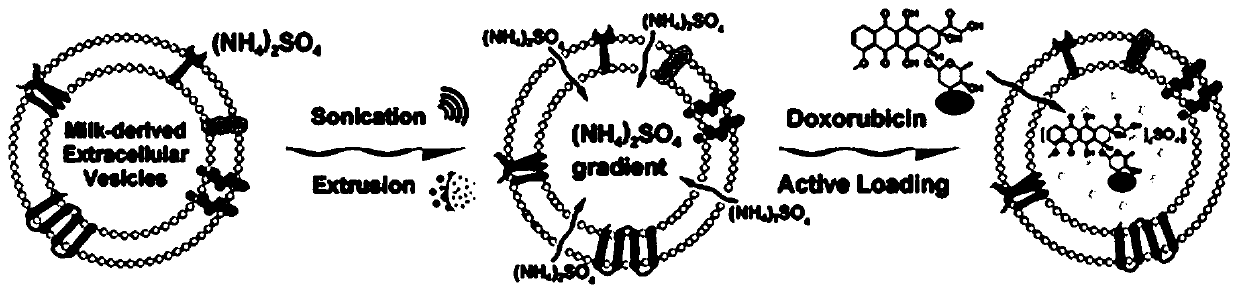
[0037] Establishment of the SEAL method:

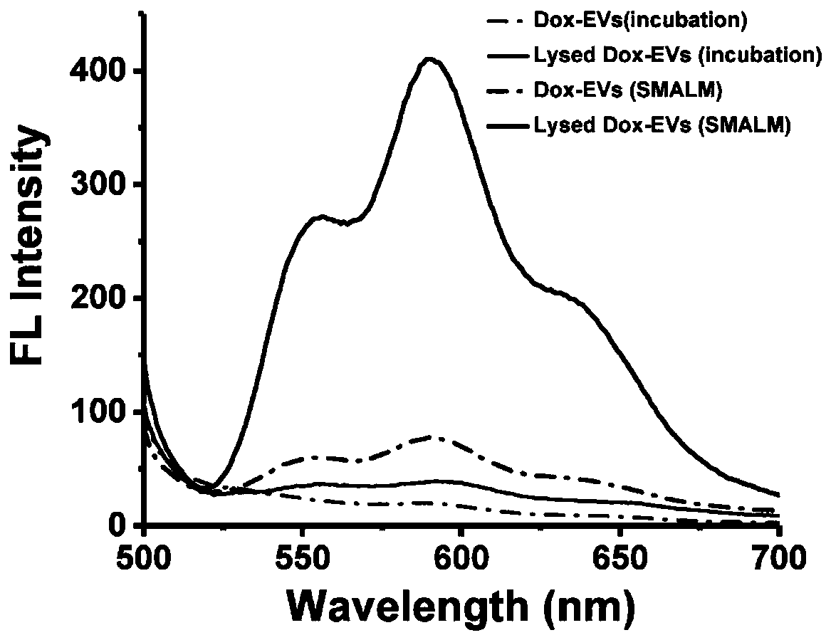
[0038] Milk exosomes (mEVs) were extracted by differential centrifugation, ultracentrifugation combined with isoelectric precipitation and membrane separation. The mEVs were resuspended in ammonium sulfate solution, placed on ice for probe sonication, and the condition of probe sonication was power 26W, 3mm tip probe, 30s on, 30s off. Then incubate at 4°C for 1 h. The incubated mEVs were extruded through a 100nm polycarbonate membrane (PC membrane) multiple times, and then dialyzed overnight with PBS to establish the ammonium ion gradient required for active drug loading. The mEVs dialyzed overnight were incubated with doxorubicin for 30 min at room temperature to obtain drug-loaded milk exosomes (Dox-mEVs). After the completion of drug loading, the excess drug in Dox-mEVs was removed by Sephadex G25 size exclusion chromatography.

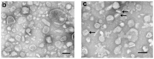
[0039] Transmission electron microscope observation:

[0040] Electron micrographs of mEVs before a...

Embodiment 2

[0056] The mEVs were resuspended in ammonium sulfate solution, placed on ice for probe sonication, and the conditions of probe sonication were power 26W, 3mm tip probe, 30s on, 30s off, 4°C, and the time was 120s. Then incubate at 4°C for 1 h. The incubated mEVs were extruded 5 times through a 100 nm polycarbonate membrane (PC membrane), and dialyzed against PBS overnight to establish the ammonium ion gradient required for active drug loading. The mEVs dialyzed overnight were incubated with mitoxantrone (MXT) for 30 min at room temperature to obtain drug-loaded milk exosomes (MXT-mEVs). After drug loading, the excess drug in MXT-mEVs was removed by Sephadex G25 size exclusion chromatography.

[0057] The content and drug loading rate of MXT were determined by UV-visible absorptiometry. First prepare MXT standard solutions with concentrations of 5 μM, 10 μM, 20 μM, 50 μM, 100 μM and 200 μM, respectively. The purified MXT-mEVs samples were added to a 96-well plate, and the ab...


PUM


Abstract

The invention relates to an extracellular vesicle active medicine loading method. The extracellular vesicle active medicine loading method comprises the following steps of performing resuspension on extracellular vesicles with an ammonium sulfate solution, performing ultrasonic treatment with a low-frequency probe, and performing incubation at 4 DEG C for 1h; repeatedly extruding the incubated extracellular vesicles through a polycarbonate membrane, performing PBS dialysis for overnight, and establishing an ammonium sulfate gradient; and performing incubation on the extracellular vesicles after dialysis for overnight and medicines, so as to obtain medicine loading dialysis extracellular vesicles. According to the method disclosed by the embodiment of the invention, the extracellular vesicle active medicine loading method based on ultrasonic treatment through the low-frequency probe and extrusion through the polycarbonate membrane can effectively improve the medicine loading rate of theextracellular vesicles and the medicine stability, is broad in application range.

Description

technical field [0001] The invention relates to the technical field of biomedicine, in particular to a method for actively loading extracellular vesicles with drugs. Background technique [0002] Extracellular vesicles (EVs), biological nanoparticles with a bilayer lipid membrane structure and a variety of biomolecules, are a novel mechanism of intercellular communication, allowing cells to exchange proteins, lipids, and genetic material. Due to their relatively small molecular structure, natural molecular transport properties, and good biocompatibility and many other advantages, extracellular vesicles have great application potential in the field of drug delivery. So far, there have been many studies to treat diseases by delivering proteins, RNA or other small molecule drugs through extracellular vesicles. [0003] At present, the drug loading methods for extracellular vesicles can be divided into two categories. One is to operate on the parental cells, that is, to transfe...

Claims


Application Information

Patent Timeline
IPC IPC(8): A61K47/46A61K9/127A61K31/704A61K31/136
CPCA61K47/46A61K9/1271A61K9/1277A61K31/704A61K31/136
Inventor 陈超翔孙梦迪林俊杰林晓洁李玉梅
Owner JIMEI UNIV
Why Eureka
Try Eureka