Mouse lung tissue primary epithelial stem cell ball culture method

A technology of stem cell spheres and culture methods, which is applied in the field of culture of primary epithelial stem cell spheres in mouse lung tissue, can solve the problems of affecting the separation effect, lack of cell surface marker morphological phenotype, and affecting the expression of cell surface antigens, so as to improve cell production. The effect of increasing the efficiency, reducing the cost of the experiment, and shortening the experiment time

Active Publication Date: 2020-08-14
JIANGSU PROVINCE HOSPITAL THE FIRST AFFILIATED HOSPITAL WITH NANJING MEDICAL UNIV
View PDF8 Cites 3 Cited by
  • Summary
  • Abstract
  • Description
  • Claims
  • Application Information

AI Technical Summary

Problems solved by technology

For the isolation of primary epithelial stem cells from lung tissue, there is currently no efficient and reliable extraction method due to the lack of specific cell surface markers and identifiable morphological phenotypes.
Some researchers isolated

Method used


Image

  • Mouse lung tissue primary epithelial stem cell ball culture method
  • Mouse lung tissue primary epithelial stem cell ball culture method
  • Mouse lung tissue primary epithelial stem cell ball culture method

Examples


Embodiment 1

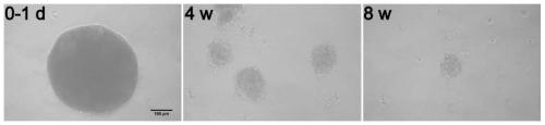
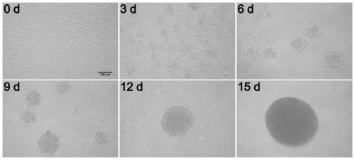
[0048] A method for culturing primary epithelial stem cell spheres of mouse lung tissue, comprising the steps of:

[0049] (1) Thoroughly disinfect the skin of the mouse with 75% alcohol, aseptically separate the lung tissue and rinse it in pre-cooled sterile PBS (phosphate buffered saline solution), remove the connective tissue and the main bronchus in the lung, and separate the lung lobes at the same time, Wash 2-3 times to remove blood.

[0050] (2) Transfer the cleaned lung lobe to a new cell culture dish with sterile tweezers, suck off the residual PBS, and use ophthalmic surgical scissors to evenly cut the lung tissue into about 1mm 3 The tissue pieces were transferred to the preheated collagenase digestion solution and digested on a constant temperature shaker (100 rpm) at 37°C for 45-60 minutes;

[0051] (3) Add an equivalent amount of DMEM / F12 medium containing 10% FBS to stop the digestion, pipette and mix well, and then filter through a 100 μm cell sieve; the filtr...


PUM


Abstract

The invention relates to a mouse lung tissue primary epithelial stem cell ball culture method. The method comprises the following steps: digesting a mouse lung lobe tissue block by adopting a preheated collagenase digestion solution, adding an equal amount of FBS-containing DMEM/ F12 culture medium to terminate digestion, extracting mouse lung tissue primary epithelial stem cells, and carrying outsuspension culture to form stem cell balls. The mouse lung tissue primary epithelial stem cell ball culture method is established through a 3D culture model, so that a lung tissue microenvironment issimulated in vitro, and an effective and reliable research model is provided for clarifying a regulation mechanism of lung epithelial stem cell proliferation and differentiation.

Description

technical field [0001] The invention belongs to the technical field of cell biology, and in particular relates to a method for culturing primary epithelial stem cell spheres of mouse lung tissue. Background technique [0002] In living organisms, almost all cells are in a complex three-dimensional (3D) structure, and their life cycle is finely regulated by the extracellular microenvironment, cell-cell and cell-extracellular matrix (ECM). The interactions form a 3D communication network that maintains tissue specificity and homeostasis. However, in the traditional two-dimensional (2D) cell adherence culture, the tissue structure and connections similar to those in vivo cannot be realized between cells, which limits the research on cell morphology, function and behavior, and also affects the research of genes and proteins. The expression, predictability of cell-based drug and toxicity screening assays have impacted. In most cases, 2D culture models do not well mimic the norm...

Claims


Application Information

Patent Timeline
IPC IPC(8): C12N5/071
CPCC12N5/0625C12N2509/00C12N2501/91C12N2501/11C12N2501/115
Inventor 周宏孔辉解卫平陈倩倩黄文
Owner JIANGSU PROVINCE HOSPITAL THE FIRST AFFILIATED HOSPITAL WITH NANJING MEDICAL UNIV
Why Eureka
Try Eureka