Double-antigen sandwich immunofluorescence chromatography kit for detecting African swine fever virus CD2v protein antibody
An African swine fever virus and double-antigen sandwich technology is applied in the field of veterinary biological products to achieve the effects of small variation coefficient, high detection sensitivity and high precision
- Summary
- Abstract
- Description
- Claims
- Application Information
AI Technical Summary
Problems solved by technology
Method used
Image

Examples
Embodiment 1
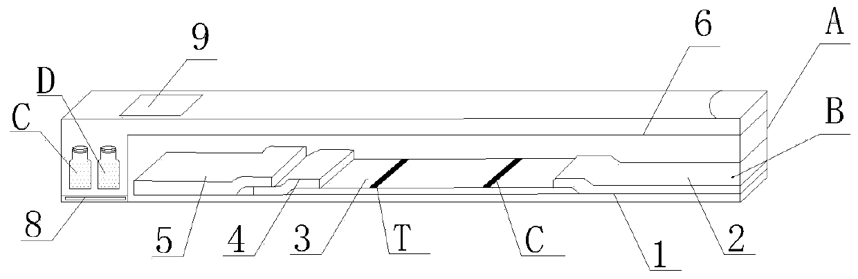
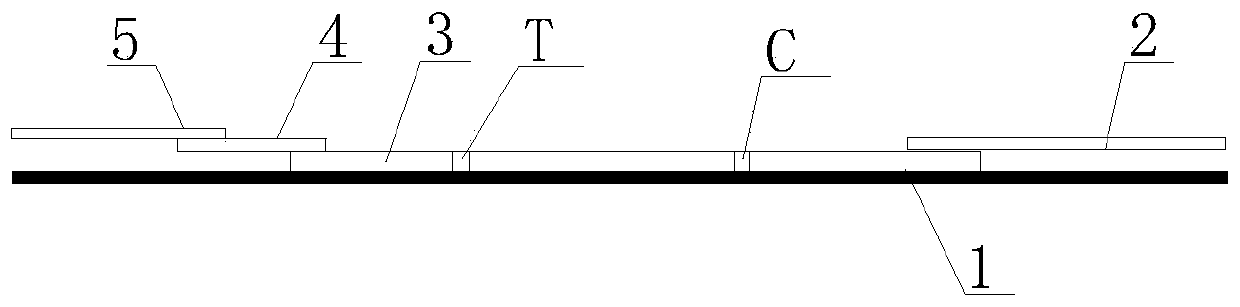
[0042] A double-antigen sandwich immunofluorescence chromatography kit for detecting African swine fever virus CD2v antibody provided by the present invention, see figure 1 , including a box body A, the box body A is equipped with a CD2v antibody immunofluorescence chromatography detection card B and a serum diluent bottle C with a serum diluent, a serum bottle D with a standard positive serum, and a standard positive serum bottle D with a standard Serum bottle E of negative serum; the CD2v antibody immunofluorescence chromatography detection card B includes a PVC bottom plate 1, and the PVC bottom plate 1 is provided with an absorption pad 2, a reaction membrane 3, a binding pad 4 and The sample pad 5, the binding pad 4 is loaded with fluorescently labeled CD2v protein and fluorescently labeled goat anti-chicken IgY, the reaction membrane 3 is provided with a detection line T and a quality control line C, the detection Line T is coated with recombinant CD2v protein, and the q...
Embodiment 2
[0046] The preparation method of the above-mentioned kit includes the following steps: flatly lay the PVC bottom plate 1 with NC film on the work surface, paste the binding pad 4, the sample pad 5 and the absorption pad 2 respectively, and cut them into 4.0mm wide strips with a strip cutter. For test strips, put each test strip into a plastic card case, place each reagent card in an aluminum film bag, add 1 pack of 1g desiccant, heat seal the seal, and it is an immunofluorescence chromatography test card.
[0047] Specifically:
[0048] 1) The coated film is made by the following method:
[0049] Dilute CD2v protein and IgY with 3% sucrose-containing PB (0.01M PH7.4) to a concentration of 1 mg / ml as the test line and quality control line solutions, draw a line on the nitrocellulose membrane, and use a marker pen at one end of the membrane Mark the quality control line and detection line. The distance between the quality control line and the detection line is 5 mm, the distanc...
PUM
 Abstract
Description
Claims
Application Information
PatSnap Eureka turns technology decisions into work you can execute. Powered by our Innovation Knowledge Graph, it runs expert workflows across engineering, life sciences, materials and intellectual property. Get your review-ready output in minutes.


