
A sample preparation method for scanning electron microscope observation of ceramic internal microstructure

A technology of ceramics and ceramic materials, which is applied in the field of scanning electron microscope sample preparation, can solve the problems of complex process, increase equipment and time cost, complex process flow, etc., and achieve the effect of simplifying process steps, saving sample preparation time, and simplifying operation process

Active Publication Date: 2022-05-10
SHANGHAI INST OF CERAMIC CHEM & TECH CHINESE ACAD OF SCI
View PDF5 Cites 0 Cited by
  • Summary
  • Abstract
  • Description
  • Claims
  • Application Information

AI Technical Summary

Problems solved by technology

At present, there are two widely used methods for observing the internal microstructure of ceramic materials by scanning electron microscopy, one is direct observation of the cross-section, and the other is observation after corroding the polished surface. The microstructure has become common knowledge in this field, but there are certain limitations and deficiencies in these two methods
[0003] First of all, using the method of directly observing the microstructure of the cross section, the observation result will be affected by the fracture mode of the material, which is not universal
For ceramic materials with intergranular fracture, the grains of the fracture surface are obvious under the scanning electron microscope, and the grain boundaries are relatively clear, which can meet the observation requirements; however, for ceramic materials with transgranular fracture, the fracture surface is flat, and there is no gap between grains and grain boundaries. Contrast, unable to meet observation requirements
[0004] The sample preparation method of corroding the polished surface, especially for ceramics with submicron grains, requires sophisticated polishing equipment and rich polishing experience. It will cause damage to the real surface of the ceramic material, and the local grains will peel off and fall off under the action of mechanical force, and the particles in the polishing solution will also remain on the surface of the sample, resulting in distortion of the internal microstructure of the sample; For samples, the surface energy is reduced, and the grain boundary area is more stable, which will bring greater difficulty to the corrosion process, and the corrosion conditions will be unstable, which will increase the cost of equipment and time
[0005] In summary, among the two methods of scanning electron microscopy to observe the internal microstructure of ceramic materials in the existing common knowledge, the first method of directly observing the fracture surface has poor versatility. The sample is not applicable; the second method of etching the polished surface has complex process, redundancy, and great uncertainty, and it is difficult to achieve fast, convenient, and accurate sample preparation requirements

Method used


Image

  • A sample preparation method for scanning electron microscope observation of ceramic internal microstructure
  • A sample preparation method for scanning electron microscope observation of ceramic internal microstructure
  • A sample preparation method for scanning electron microscope observation of ceramic internal microstructure

Examples


Embodiment 1

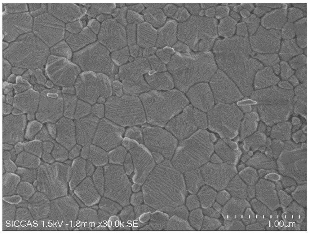
[0040] Example 1: Preparation of scanning electron microscope images of hot corrosion cross-sections of magnesium aluminum spinel ceramics

[0041] Using commercial magnesia-aluminum spinel powder as raw material, the ceramic green body with a relative density of 56% was prepared by injection molding method, and sintered in an air atmosphere at 1500°C for 6 hours to obtain a magnesia-alumina spinel ceramic with a relative density of 93%.

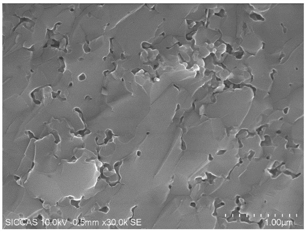
[0042] Break the obtained magnesia-aluminum spinel ceramics, and take a fresh and clean section as a sample. For the microstructure diagram of the fresh section, see figure 2 , it can be seen from the figure that there is no contrast between grains and grain boundaries, and the ceramic sample fractures in the form of transgranular fracture;

[0043] Place the sample in a muffle furnace for thermal corrosion, control the corrosion temperature to 1200°C, and hold the temperature for 3 hours;

[0044] Coating treatment (material chromium, thi...

Embodiment 2

[0046] Example 2: Preparation of scanning electron microscope images of hot corrosion cross-sections of alumina ceramics

[0047] Using commercial alumina powder as raw material, a ceramic green body with a relative density of 53% was prepared by injection molding;

[0048] The alumina ceramic green body was sintered at 1400°C for 3 hours to obtain alumina ceramics with a relative density of 85%. The sintered ceramic samples were crushed, and fresh and clean sections were taken as samples;

[0049] Put the sample in a muffle furnace for thermal corrosion, the corrosion temperature is 1000 ° C, and the holding time is 3 hours;

[0050] Coating treatment (material chromium, thickness <5nm) is performed on the sample after thermal corrosion to enhance the conductivity of the ceramic sample and avoid charging phenomenon during scanning electron microscope observation.

[0051] Place the coated sample on the sample stage of the scanning electron microscope, put it into the sample ...

Embodiment 3

[0052] Example 3: Preparation of scanning electron micrographs of acid corrosion cross-section of alumina ceramics

[0053] Using commercial alumina powder as raw material, a ceramic green body with a relative density of 56% was prepared by injection molding;

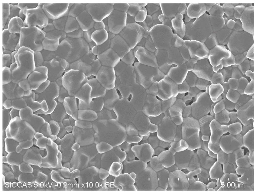
[0054] The alumina ceramic green body was sintered at 1550°C for 6 hours to obtain alumina ceramics with a relative density of 95%. The sintered ceramic samples were crushed and a fresh and clean section was taken as a sample; the microstructure diagram of the fresh section can be found in Figure 5 , it can be seen from the figure that there is no contrast between grains and grain boundaries in some areas, and only grain boundaries in some areas are clear. The ceramic sample fractures in a partial transgranular fracture mode;

[0055] Place the sample in phosphoric acid (concentration 15mol / L) for corrosion, the corrosion temperature is 300°C, and the holding time is 30 minutes;

[0056] Coating treatment (material go...


PUM

PropertyMeasurementUnit
particle sizeaaaaaaaaaa
thicknessaaaaaaaaaa

Abstract

The invention relates to a sample preparation method for observing the internal microstructure of ceramics with a scanning electron microscope. A ceramic sample with submicron grains is broken to obtain a fresh section, and then subjected to thermal corrosion or acid corrosion to obtain a ceramic material to be observed; The fracture mode of the ceramic sample with submicron grains is complete transgranular fracture or partial transgranular fracture.

Description

technical field [0001] The invention relates to a sample preparation method for observing the internal microstructure of ceramics with a scanning electron microscope, in particular to a method for preparing a scanning electron microscope sample of a ceramic material with submicron grains, and belongs to the technical field of material testing and analysis. Background technique [0002] The grain morphology, size, and distribution of ceramic materials will have a significant impact on the mechanical, optical, and electrical properties of the material. Therefore, clear imaging of the microstructure of ceramic materials is a prerequisite for characterizing the microscopic properties of ceramics. At present, there are two widely used methods for observing the internal microstructure of ceramic materials by scanning electron microscopy, one is direct observation of the cross-section, and the other is observation after corroding the polished surface. Microstructure has become comm...

Claims


Application Information

Patent Timeline
Patent Type & Authority Patents(China)
IPC IPC(8): G01N23/2202
CPCG01N23/2202
Inventor 章健刘梦玮赵瑾岛井骏藏王士维陈晗陈鹤拓
Owner SHANGHAI INST OF CERAMIC CHEM & TECH CHINESE ACAD OF SCI
Learn More