EpCAM gene expression detection kit

A detection kit and gene expression technology, applied in the field of molecular biology, can solve the problem of difficult control of factors such as limited sample sources and the accuracy of test results

Pending Publication Date: 2020-10-23
SUREXAM BIO TECH
View PDF3 Cites 2 Cited by
  • Summary
  • Abstract
  • Description
  • Claims
  • Application Information

AI Technical Summary

Problems solved by technology

[0006] At present, immunohistochemistry and real-time quantitative PCR are mostly used in the detection of EpCAM expression. These two methods have certain limitations in actual detection, including the limited source of samples and many factors that affect the accuracy of detection results, which are difficult to control.

Method used


Image

  • EpCAM gene expression detection kit
  • EpCAM gene expression detection kit
  • EpCAM gene expression detection kit

Examples


Embodiment 1

[0048] Example 1 EpCAM gene expression detection kit

[0049] The EpCAM gene expression detection kit (in situ hybridization method) described in the present embodiment mainly includes:

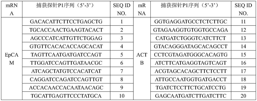
[0050] 1. Capture Probes

[0051] From the 5' end to the 3' end of the capture probe is a specific P1 sequence capable of binding to the target mRNA to be detected, a solubilizing group, and a P2 sequence capable of binding to the P3 sequence of the amplification probe, aiming at the same target mRNA The P2 sequence in the capture probe is the same; the specific P1 sequence is a peptide nucleic acid sequence with a base length of 16-20bp; the solubilizing group is selected from O-linker, E-linker, X-linker At least one: the P2 sequence is a sequence that does not have a hairpin structure, does not form dimers or mismatches within and between probes, and does not specifically bind to P1 and EpCAM gene mRNA. The solubilizing group can improve the solubility of the capture probe, and at the sa...

Embodiment 2

[0077] Example 2 Using the kit described in Example 1 to detect the sample

[0078] The formula of described various solutions is as table 6:

[0079] Table 6

[0080]

[0081] In this embodiment, the blood samples of tumor patients are preferred, and the expression level of EpCAM gene of circulating tumor cells in the samples is detected, wherein the capture probe mixture, the amplification probe mixture, and the chromogenic probe mixture all use the reagents described in Example 1 All probes in the box corresponding to the list.

[0082] 1. Extract 5ml of blood from the patient's vein into a vacuum blood collection tube to obtain a blood sample

[0083] 2. Sample pre-treatment, the cells to be tested are filtered onto the filter membrane

[0084] (1) Transfer 5ml of blood sample to a centrifuge tube containing 10ml of erythrocyte lysate, shake and mix well for 10 seconds, centrifuge at 600×g for 5 minutes, discard the supernatant to keep the cell pellet, and resuspend ...

Embodiment 3

[0132] Embodiment 3 The impact of different types of capture probes on the detection effect of the kit

[0133] In order to evaluate the detection effect of kits composed of different types of capture probes, experimental groups 1-2 were designed. Except for the different types of capture probes, the other components of the two groups were the same. The specific design is shown in Table 9.

[0134] Table 9 Selection of kit capture probes

[0135] test group capture probe type Experimental group 1 Capture probes of the present invention Experimental group 2 Traditional Linear Oligonucleotide Probes

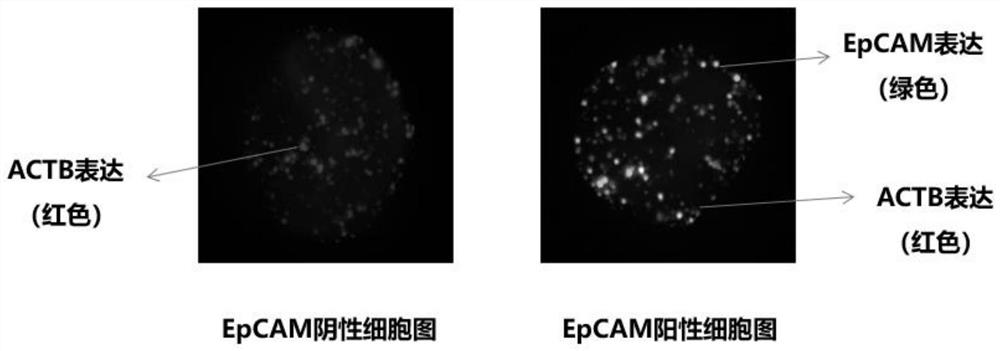
[0136] Using the kit designed and prepared above, the blood samples of 10 tumor patients (numbered 1-10) were detected according to the detection process and method described in Example 2, and the cells with DAPI blue fluorescent signal in each sample were read. , count the number of cells expressing green / red fluorescence, and the cells expressing t...


PUM

PropertyMeasurementUnit
concentrationaaaaaaaaaa

Abstract

The invention provides an EpCAM gene expression detection kit. The kit comprises a capture probe for detecting EpCAM gene mRNA and a signal amplification system; the signal amplification system comprises amplification probes and labeling probes; the capture probes sequentially comprise a specific P1 sequence which can be combined with mRNA of an EpCAM gene to be detected, a solubilizing group anda P2 sequence from the 5' end to the 3' end, the specific P1 sequence, and the specific P1 sequence is a peptide nucleic acid sequence with the length of 16-20 bp; the amplification probes are connected to the capture probe and the labeling probes, and bases of each amplification probe from the 5' end to the 3' end are sequentially composed of a P3 sequence, a spacer arm sequence and a P4 sequencewhich can be complementarily paired with the P2 sequence; and the labeling probes are connected to the amplification probes and fluorophore, each labeling probe has a P5 sequence complementarily paired with the corresponding amplification probe P4, and the tail end of each labeling probe is modified with the fluorophore. Through optimization of the detection probes, the kit has the advantages ofhigh accuracy, short detection time, time saving, labor saving and the like.

Description

technical field [0001] The invention belongs to the technical field of molecular biology, and in particular relates to an EpCAM gene expression detection kit. Background technique [0002] Epithelial cell adhesion molecule (EpCAM), also known as CD326, was originally described as a dominant surface antigen in human colon cancer, a transmembrane glycoprotein that mediates epithelial-specific intercellular adhesion, and its gene is located on human chromosome 2p21 , containing 9 exons. EpCAM is expressed in most normal epithelial cells and malignant epithelial tumor cells such as gastrointestinal cancer, but its expression level is different in different cell types and organs. EpCAM expression is strongly positive in most epithelial cell types throughout the body, and is mainly concentrated in the lateral membrane and basement membrane, but in non-epithelial tissues such as lymphoid-derived and myeloid-derived cells, mesenchymal types, muscle or neuroendocrine tissues not ex...

Claims


Application Information

Patent Timeline
Patent Type & Authority Applications(China)
IPC IPC(8): C12Q1/6886C12Q1/6841
CPCC12Q1/6886C12Q1/6841C12Q2600/158C12Q2600/166C12Q2525/107C12Q2565/519C12Q2563/107
Inventor 许嘉森吴诗扬黄洁芬刘志明刘芳
Owner SUREXAM BIO TECH
Why Eureka
Try Eureka