Method for monitoring temperature of lesion area in real time through magnetic resonance and application thereof

A technology of real-time monitoring and lesion area, applied in contraceptives, heating appliances for treatment, cooling appliances for treatment, etc., can solve problems such as inaccurate temperature measurement and wrong treatment evaluation, and achieve high safety and reduce Trauma, high-resolution effects

Active Publication Date: 2020-10-27
HARBIN MEDICAL UNIVERSITY
View PDF7 Cites 1 Cited by
  • Summary
  • Abstract
  • Description
  • Claims
  • Application Information

AI Technical Summary

Problems solved by technology

However, the temperature accuracy obtained by MRI temperature measurement is ±1°C. When the commonly used PRF (proton nuclear magnetic resonance frequency) phase subtraction method is used for in vivo real-time temperature measurement, it is easily affected by fat signals, motion artifacts, and phase windi

Method used


Image

  • Method for monitoring temperature of lesion area in real time through magnetic resonance and application thereof
  • Method for monitoring temperature of lesion area in real time through magnetic resonance and application thereof

Examples


Embodiment 1

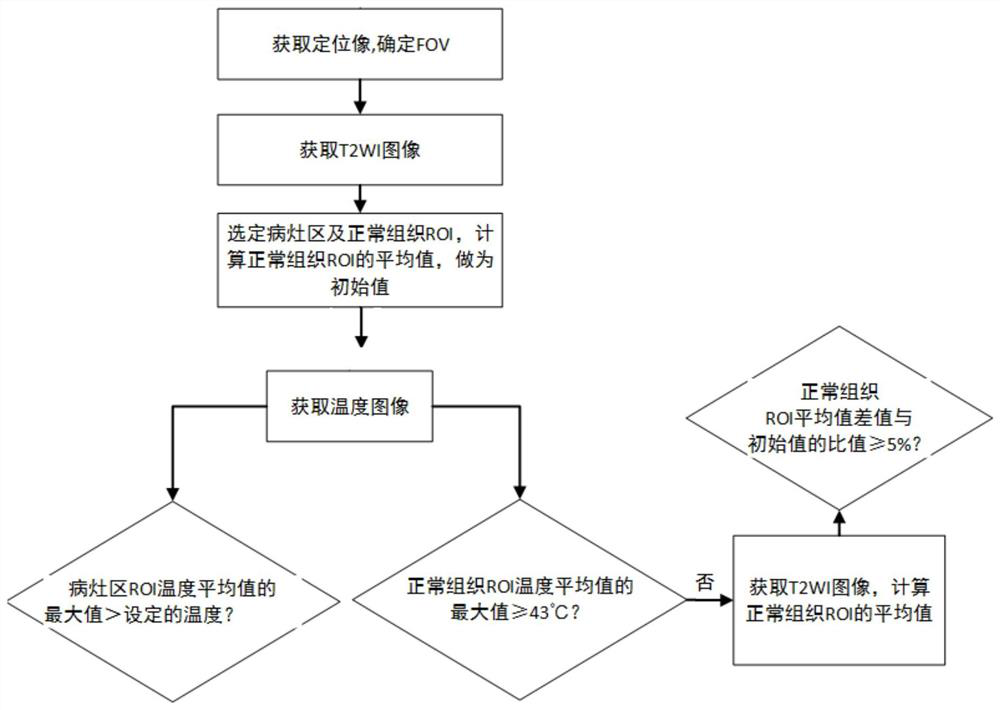
[0032] Such as figure 1 As shown, a method for magnetic resonance real-time monitoring of the temperature of the lesion area includes the following steps:

[0033] (1) Obtain positioning images: Send the lesion area into the center of the MRI magnet, use the positioning image sequence to obtain low-resolution positioning images of the lesion area, and determine the field of view (FOV) of T2WI images and temperature imaging based on the positioning images;

[0034] Preferably, the gradient echo family sequence is used to obtain the positioning image, for example, the FLASH sequence is used to quickly collect data to obtain a low-resolution positioning image.

[0035] (2) Obtaining T2WI images: based on the FOV described in step (1), using T2WI sequences to obtain T2WI images of high-resolution anatomical structures of the lesion area and surrounding normal tissues of the lesion area and statistically analyze them;

[0036] Preferably, T2WI images are collected using spin echo-ec...

Embodiment 2

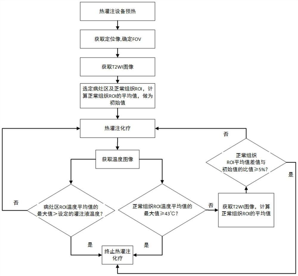
[0043] In this embodiment, a GE 3.0T MR imager is used to guide patients with bladder cancer in real time to receive pirarubicin hydrochloride hyperthermic perfusion chemotherapy. Routine clinical 1.5T and 3.0T MR imagers are available, such as uMR 3.0T MR imager, GE 3.0T MR imager, Philips Achieva 1.5T MR imager, etc.

[0044] The application of a method of magnetic resonance real-time monitoring of the temperature of the lesion area described in Example 1 in real-time guidance of hyperthermic perfusion chemotherapy for bladder cancer, such as figure 2 As shown, the specific steps are as follows:

[0045] Step 1: Prepare, check, and connect relevant perfusion chemotherapy equipment according to the routine process of hyperthermic perfusion chemotherapy, connect the one-time-use body cavity hyperthermic perfusion therapy pipeline components, and preheat the equipment. Prepare perfusion fluid, patient-related preparation, no need to insert temperature sensor in patient perfus...


PUM


Abstract

The invention provides a method for monitoring the temperature of a lesion area in real time through magnetic resonance and application thereof. The temperature average value of an ROI of an MRI temperature distribution diagram and a signal average value of a T2WI image are integrated to serve as judgment standards, and the situation that the temperature monitoring effect is affected due to the fact that calculation of the ROI temperature, the signal average value and the like is inaccurate due to the fact that a single temperature imaging parameter is easily affected by external factors is avoided. The method can be applied to real-time guidance of bladder cancer hyperthermia perfusion chemotherapy; the temperature change in the hyperthermia perfusion chemotherapy process is evaluated according to the ROI average signal intensity change condition, and whether hyperthermia perfusion chemotherapy damages normal tissues or not can be observed in real time by comparing the average value of the perfusion region temperature distribution diagram and the T2WI image ROI with an initial value.

Description

technical field [0001] The invention relates to the field of magnetic resonance (MRI) application technology, in particular to a method for real-time monitoring of the temperature of a lesion area by magnetic resonance and its application. Background technique [0002] The existing temperature measurement methods for the lesion area are mainly based on the sensors inserted in the lesion area. The range and accuracy of temperature measurement are greatly affected by the number and distribution of temperature sensors, and not all the temperature of the lesion area is obtained, let alone the temperature around the lesion area. Therefore, it is of great significance to accurately and effectively obtain the temperature of the lesion area and its surrounding normal tissues, and use it in hyperthermic perfusion chemotherapy, microwave ablation, ultrasound focusing, and radiofrequency ablation. [0003] Hyperthermic perfusion chemotherapy is a way to treat bladder cancer. A large-ca...

Claims


Application Information

Patent Timeline
IPC IPC(8): G06T7/00G06T7/70A61F7/12A61F7/00
CPCG06T7/0012G06T7/70A61F7/12A61F7/0053A61F2007/0059G06T2207/10088G06T2207/20104
Inventor 孙夕林王洪斌杨春升王凯徐佐宇杨丽丽吴丽娜官小红
Owner HARBIN MEDICAL UNIVERSITY
Why Eureka
Try Eureka