Rift valley fever virus humanized monoclonal antibody and application thereof

一种抗体、抗原的技术,应用在医药领域,能够解决临床数据不多、成本问题、不可能大范围接种等问题,达到高应用价值、预防感染的效果

Active Publication Date: 2020-11-24
INST OF MICROBIOLOGY - CHINESE ACAD OF SCI
View PDF7 Cites 3 Cited by
  • Summary
  • Abstract
  • Description
  • Claims
  • Application Information

AI Technical Summary

Problems solved by technology

In addition, high-risk groups in Africa can be vaccinated with inactivated formalin vaccine, but due to cost issues, it is impossible to vaccinate on a large scale
After people get sick, ribavirin may have a therapeutic effect, but there are not many clinical data at present

Method used


Image

  • Rift valley fever virus humanized monoclonal antibody and application thereof
  • Rift valley fever virus humanized monoclonal antibody and application thereof
  • Rift valley fever virus humanized monoclonal antibody and application thereof

Examples


Embodiment 1

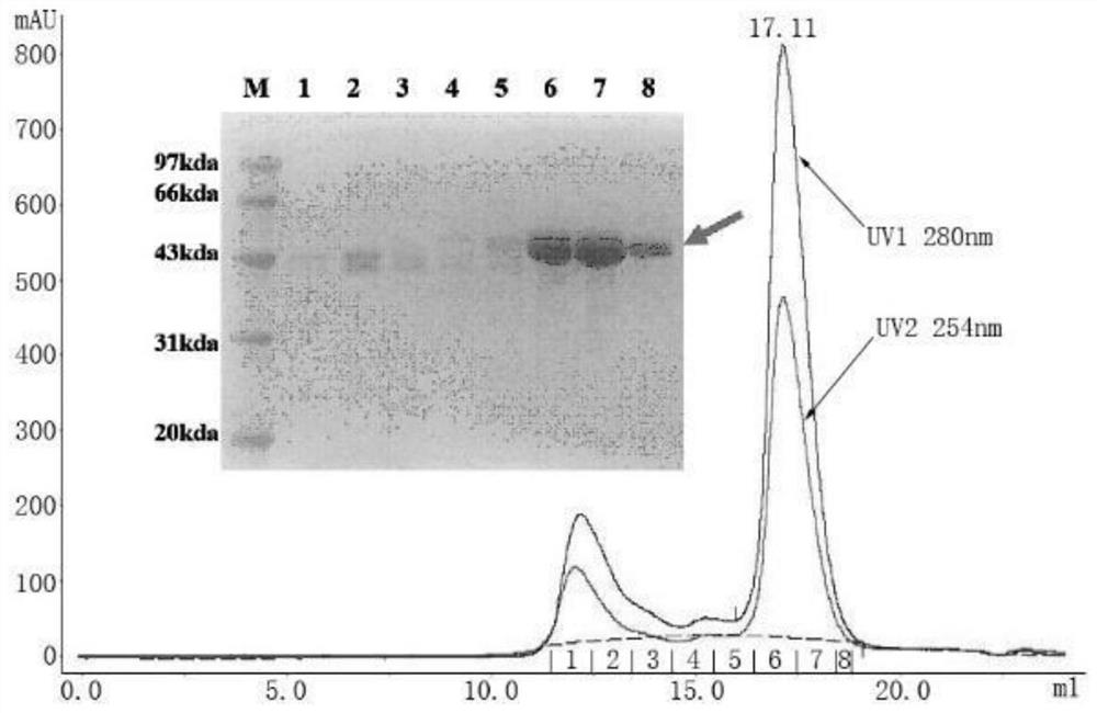
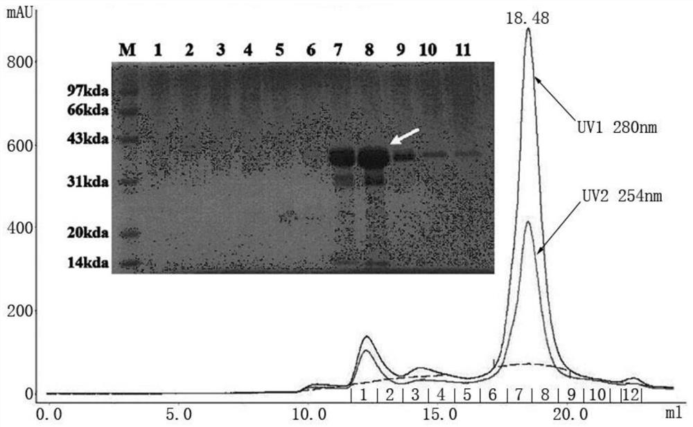
[0069] Example 1: Expression and purification of RVFV Gn and Gc proteins

[0070] The genes encoding the RVFV Gn (amino acid sequence shown in SEQ ID NO:23) and Gc (amino acid sequence shown in SEQ ID NO:24) proteins were respectively connected to the pFastBac1 vector, wherein Gn was connected to the N-terminal of the Gc protein gene There is an insect cell membrane protein Gp67 signal peptide sequence for antibody secretion; a His tag is attached to the C-terminus, which is convenient for purification and staining of B cells; all coding genes have been optimized for insect cell codons.

[0071] Transform Escherichia coli DH10Bac competent cells with the pFastBac1 vector containing the target gene, and perform blue-white screening of recombinant baculoviruses on a plate containing ampicillin, kanamycin, tetracycline and Blue-gal. After the white plaques were identified by PCR with the upstream and downstream primers of M13, the positive clones were shaken, and the recombinant ...

Embodiment 2

[0075] Example 2: Isolation of RVFV Gn and Gc protein-specific memory B cells

[0076] With the patient's informed consent, 30 mL of blood was collected and PBMCs were isolated.

[0077] Separated PBMCs in 10 7 Individual / mL density and RVFV Gn and RVFV Gc proteins with a final concentration of 100nM were incubated on ice for half an hour, then washed twice with PBS, and then incubated with the following antibodies on ice for half an hour: anti-human CD3 / PE-Cy5, anti-human CD16 / PE-Cy5, anti-human CD235a / PE-Cy5, anti-human CD19 / APC-Cy7, anti-human CD27 / Pacific Blue, anti-human CD38 / APC, anti-human IgG / FITC and anti -His / PE, then washed 2 times with PBS.

[0078] The PE-Cy5-APC-APC-Cy7+Pacific Blue+FITC+PE+ cells were collected by FACSAria III sorting, and directly collected into a 96-well plate, 1 cell / well.

Embodiment 3

[0079] Example 3: Single B cell PCR and sequence analysis

[0080] The cells obtained in Example 2 were reverse-transcribed by Superscript III reverse transcriptase (Invitrogen), and reacted at 55° C. for 60 min.

[0081] Using this reverse transcription product as a template, PCR was performed with HotStar Tap Plus enzyme (QIAgen) to amplify the antibody variable region sequence (PCRa). The reaction conditions were as follows: 95°C, 5min; 95°C, 30s, 55°C (heavy chain / κ chain) / 50°C (λ chain), 30s, 72°C, 90s, 35 cycles, 72°C, 7min.

[0082] Use this as a template for another round of PCR (PCRb), the conditions are as follows: 95°C, 5min; 95°C, 30s, 58°C (heavy chain) / 60°C (κ chain) / 64°C (λ chain), 30s, 72 ℃, 90s, 35 cycles, 72℃, 7min.

[0083] 1.2% agarose gel electrophoresis to separate the PCR products. The bands with a size of 400-500 bp were recovered and sent to the sequencing company for sequencing. The sequencing results were analyzed with IMGT online software.

[00...


PUM


Abstract

The invention discloses a rift valley fever virus humanized monoclonal antibody and an application thereof, and belongs to the technical field of medicines. According to the invention, RVFV glycoprotein Gn and Gc expressed by rod-shaped viruses are used as antigens; memory B cells capable of being combined with the RVFV glycoprotein Gn and Gc are screened from PBMCs of a rift valley fever rehabilitation patient; and through experiments of single B cell sequencing, in-vitro neutralization, in-vivo protection and the like, eight strains of Gn humanized monoclonal antibodies capable of efficiently neutralizing RVFV infection and a strain of a Gc humanized monoclonal antibody are identified. The humanized monoclonal antibody disclosed by the invention has extremely high in-vitro neutralizationactivity (IC50 can be as low as 1.93 + / - 0.6 pM, reaching a pM level), can effectively treat mice infected by RVFV and prevent infection of RVFV on the mice, and has extremely high application valuein clinical treatment and prevention of RVFV infection.

Description

technical field [0001] The invention relates to a Rift Valley fever virus human monoclonal antibody and application thereof, belonging to the technical field of medicine. Background technique [0002] Rift Valley fever virus (RVFV) is one of the arboviruses that seriously threaten human and animal health. It can cause the zoonotic Rift Valley fever (Rift Valley fever, RVF), causing livestock death and abortion. Its surface is covered with envelopes and glycoprotein protrusions, and the viral genome (S, M, and L) is composed of 3 segments of negative-strand RNA. Among them, the M segment encodes surface glycoproteins Gn and Gc, and encodes non-structural proteins at the 5' end of Gn Protein (NSm), and Gn and Gc are the main envelope proteins of the virus, which are the key proteins for virus invasion and membrane fusion; its spread is very wide, and sheep, goats, cattle, buffaloes, camels and other livestock can be infected with the virus ; Its lethality rate is very high, ...

Claims


Application Information

Patent Timeline
Patent Type & Authority Applications(China)
IPC IPC(8): C07K16/10C12N15/13C12N15/866C12N5/10A61K39/42A61P31/14
CPCC07K16/10A61P31/14C07K2317/56C07K2317/52A61K2039/505C07K2317/24C07K2317/92C07K2317/76Y02A50/30
Inventor 严景华王奇慧马桐杨化冰高福马素芳
Owner INST OF MICROBIOLOGY - CHINESE ACAD OF SCI
Why Eureka
Try Eureka