Method for establishing mouse model with multiple myeloma combined with chronic graft-versus-host disease
A multiple myeloma and establishment method technology, applied in the field of establishment of multiple myeloma combined with chronic graft-versus-host disease mouse model, to achieve the effects of good repeatability, easy acquisition, and simple process
- Summary
- Abstract
- Description
- Claims
- Application Information
AI Technical Summary
Problems solved by technology
Method used
Image
Examples
Embodiment 1
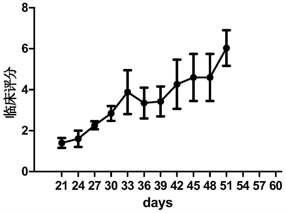
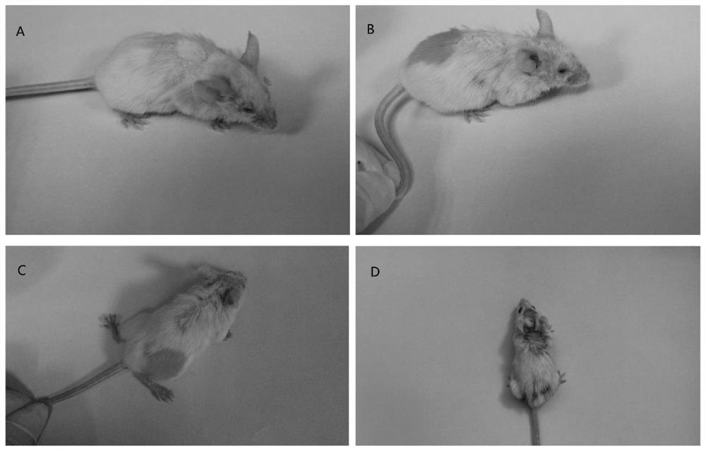
[0025] This example is an example of establishing the mouse multiple myeloma combined with chronic graft-versus-host disease model of the present invention, and the operation is as follows.
[0026] 1. Mouse preparation
[0027] Specific pathogen Free (SPF) grade NCG (NOD-SCID-IL2rg - / - ) 8-week-old male mice (female mice can also be used) as model mice. The mice in this example were purchased from the Model Animal Research Institute of Nanjing University. The rats were randomly divided into 6 groups, 5 in each group; among them:
[0028] Group A (normal control group): feed synchronously, without intervention;
[0029] Group B (multiple myeloma control group): after whole body irradiation of 100cGy, the suspension of multiple myeloma luciferase reporter gene (Luciferase) stably transfected cell line (IM-9) was injected into NCG mice; Tail vein injection of PRMI1640 culture solution;
[0030] Group C (multiple myeloma+GFP-T control group): After whole-body irradiation of ...
Embodiment 2
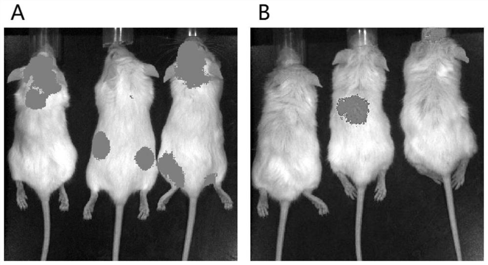
[0052] This example is an example of in vivo imaging of tumor burden in mice with multiple myeloma combined with cGVHD.
[0053] 1. For IM-9 cells, due to the expression of luciferase, imaging can be performed through the substrate to detect the residual and growth status of IM-9 cells;
[0054] 2. Inject the substrate D-luciferin 200ul (15mg / ml) through the intraperitoneal cavity, and after 5 minutes of timing, put it into the anesthesia room attached to the in vivo imager, and perform anesthesia with isoflurane at the same time;
[0055] 3. Put the anesthetized mouse into the imager for imaging, and perform imaging in the automatic exposure mode (that is, the exposure time will be automatically adjusted according to the signal strength);
[0056] 4. After imaging, put the mice back into the cage for feeding;
[0057] 5. Data analysis.
[0058] like image 3 Shown: A is the in vivo imaging of mice on the 7th day after the tumor was planted, and all the tumors in the mice w...
Embodiment 3
[0060] This example is an example of flow cytometry detection of tumor burden in mice with multiple myeloma complicated with cGVHD.
[0061] 1. For the IM-9 cell line, because it expresses the CS1 antigen, the expression of the CS1 antigen in the peripheral blood can be detected by flow cytometry, and the residual situation of the IM-9 cells can be detected;
[0062] 2. Collect blood from tail vein of mice in multiple myeloma+CS1CAR-T group;
[0063] 3. Stain the peripheral blood of the mouse with anti-human CS1 for 30 minutes. After 30 minutes, add 500ul of erythrocyte lysate into the flow tube, lyse for 10 minutes, add 3ml of PBS to wash, centrifuge at 300g for 5min, wash with PBS again, add 200ul PBS spare.
[0064] 4. Detect the expression of CS1 antigen on the computer, and analyze the data.
[0065] like Figure 4 Shown: the expression of CS1 antibody in the mice of group D, CS1 in the figure further proves the tumor burden of the mice, and the residual multiple myelo...
PUM
 Abstract
Description
Claims
Application Information
 - R&D
- Intellectual Property
- Life Sciences
- Materials
- Tech Scout
- Unparalleled Data Quality
- Higher Quality Content
- 60% Fewer Hallucinations
Browse by: Latest US Patents, China's latest patents, Technical Efficacy Thesaurus, Application Domain, Technology Topic, Popular Technical Reports.
© 2025 PatSnap. All rights reserved.Legal|Privacy policy|Modern Slavery Act Transparency Statement|Sitemap|About US| Contact US: help@patsnap.com


