Method for measuring tissue distribution of MSC

A technology of tissue distribution and real-time fluorescence quantification, which is applied in the field of biomedical detection, can solve the problem of inability to accurately quantify the number of mesenchymal stem cells in tissues, and achieve the effects of low detection limit, accurate quantification and high specificity

Pending Publication Date: 2021-06-29
山东省齐鲁细胞治疗工程技术有限公司 +1
View PDF0 Cites 1 Cited by
  • Summary
  • Abstract
  • Description
  • Claims
  • Application Information

AI Technical Summary

Problems solved by technology

[0003] Aiming at the problem that the number of mesenchymal stem cells in tissues cannot be accurately quantified in the prior art, the present invention provides a method for quantitatively measuring the tissue distribution of MSCs, using real-time fluorescent quantitative PCR

Method used


Image

  • Method for measuring tissue distribution of MSC
  • Method for measuring tissue distribution of MSC
  • Method for measuring tissue distribution of MSC

Examples


Embodiment 1

[0023] Example 1 Determination of detection primers

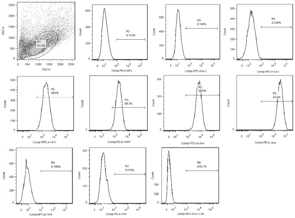
[0024] (1) Will figure 1 1×10 human umbilical cord-derived mesenchymal stem cells with positive rates of CD73, CD105, CD44, and CD90 detected by flow cytometry above 98%, and negative rates of CD34, CD45, and HLA-DR below 0.2% 7 For each, use blood / cell / tissue genomic DNA extraction kit (DP304, Tiangen Biochemical Technology (Beijing) Co., Ltd.) to extract DNA, calculate the total amount of DNA, and then half-dilute 9 times to obtain 10 serial concentrations of DNA standards solution;

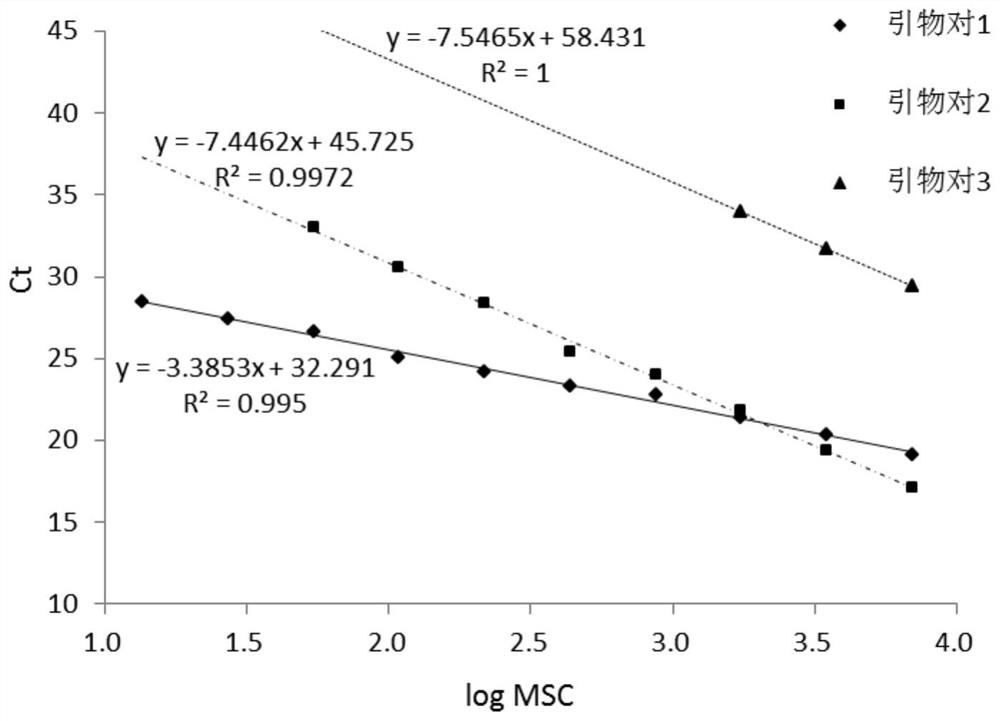
[0025] (2) According to the sequence of SEQ ID NO:7-9, design three pairs of primers as shown in the table below and synthesize them:

[0026]

[0027] Use the DNA standard solution in step (1) as a template to perform real-time fluorescent quantitative PCR, and the reaction system is as follows:

[0028]

[0029] The amplification reaction is as follows:

[0030]

[0031] The melting curve acquisition reaction is as follows: 95...

Embodiment 2

[0034] Example 2 Repeatability detection

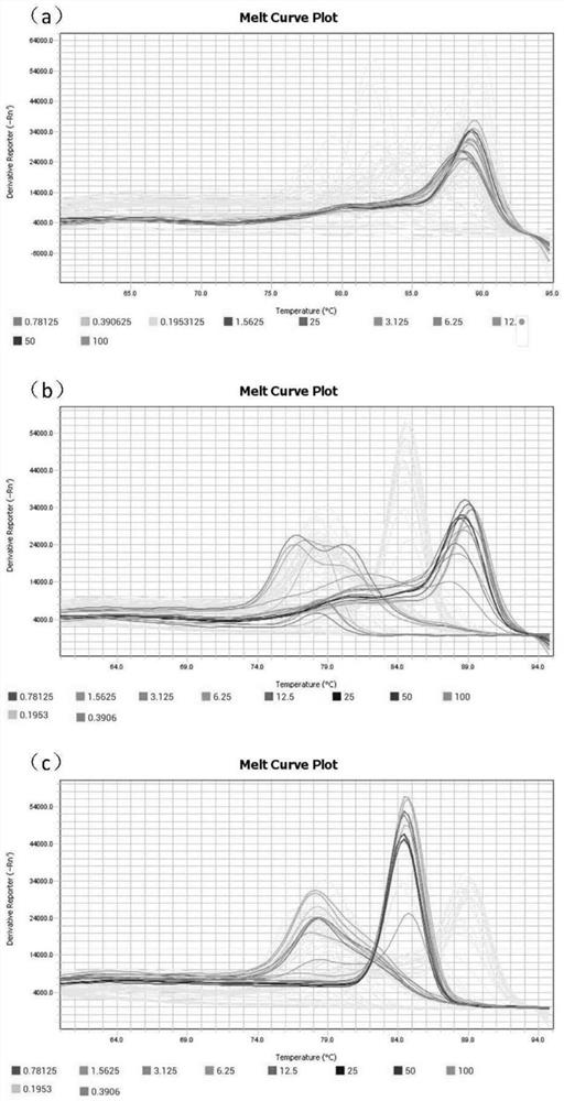
[0035] The first pair of primers was tested for repeatability, and the DNA standard solutions of 10 serial concentrations in Example 1 were also taken, and the amplification experiment was carried out according to the method in Example 1, and the melting curve obtained was as follows: Figure 4 As shown, it can be seen from the melting curve that primer pair 1 has no non-specific amplification, which can be used to evaluate the method, and the coefficient of variation (CV) is calculated according to the Ct value of each standard solution. When the amount of DNA changes from 0.19ng to 100ng, the coefficient of variation is from 0.97% to 2.76%, which is less than 3%, which can be used for subsequent quantitative detection.

Embodiment 3

[0036] Example 3 Quantitative detection of tissue distribution of mesenchymal stem cells

[0037] (1) Will figure 1 The positive rates of CD73, CD105, CD44, and CD90 detected by flow cytometry are above 98%, and the negative rates of CD34, CD45, and HLA-DR are below 0.2%. The cell density in the liquid is 1×10 7 cells / mL, BALB / c-nude nude mice (female mice) were used as test subjects, injected into the tail vein, each injection volume: 200μL, 15 mice in total;

[0038] At 24h, 48h, 72h, 96h, and 120h after injection, 3 nude mice were randomly selected at each time point and killed by necking after anesthesia. The heart, liver, spleen, lung, and kidney tissues were collected, washed with blood, sucked water, and weighed. Then freeze and store at -80°C;

[0039] After all the samples were taken, DNA was extracted using the Blood / Cell / Tissue Genomic DNA Extraction Kit (DP304, Tiangen Biochemical Technology (Beijing) Co., Ltd.);

[0040] (2) Using the sequence shown in SEQ ID ...


PUM


Abstract

The invention provides a method for determining tissue distribution of MSC. The method comprises the following steps of (1) infusing human mesenchymal stem cells into a laboratory mouse body, sampling organs or tissues to be monitored at regular intervals, and extracting DNA (deoxyribonucleic acid) samples; (2) extracting DNA from the determined number of human mesenchymal stem cells and diluting the DNA into DNA solutions with a series of concentrations; (3) carrying out real-time fluorescent quantitative PCR (Polymerase Chain Reaction) by taking the sequences as shown in SEQ ID NO: 1 and SEQ ID NO: 2 as primers and taking the DNA sample in the step (1) and the DNA solution in the step (2) as templates; and (4) making a standard curve according to the CT values of the DNA solutions with the series concentrations in the step (2), and calculating the number of the DNA samples in the step (1). According to absolute quantification of the tissue mesenchymal stem cells, the colonization rate of the mesenchymal stem cells in each tissue and the metabolism condition of the mesenchymal stem cells in each tissue along with time in the body can be detected, and a reference basis is provided for designing the administration route and dosage in a clinical scheme.

Description

technical field [0001] The invention belongs to the technical field of biomedical detection, and in particular relates to a method for quantitatively measuring MSC tissue distribution. Background technique [0002] Mesenchymal stem cells are a kind of pluripotent stem cells derived from mesoderm, which have good self-renewal and multidirectional differentiation capabilities. Because of its wide range of sources, multi-directional differentiation potential and strong homing ability, it has been well applied in clinical treatment. The declaration of stem cells as a drug needs to rely on animals for preclinical research, especially the research on drug level effectiveness and drug metabolism level. At present, the use of isotope tracer, small animal live imaging, common PCR and other technologies can realize the in vivo or in vitro tracer of cells. Accurate quantification of cell content. Contents of the invention [0003] In view of the problem that the number of mesenchy...

Claims


Application Information

Patent Timeline
Patent Type & Authority Applications(China)
IPC IPC(8): C12Q1/6851C12Q1/6888
CPCC12Q1/6851C12Q1/6888C12Q2531/113C12Q2545/114C12Q2563/107
Inventor 曲纳温丁可韩镇谭毅马贺然
Owner 山东省齐鲁细胞治疗工程技术有限公司
Why Eureka
Try Eureka