A kind of nanometer fluorescent probe solution and application of hepatic cell labeling below freezing point

A nano-fluorescence probe and nano-fluorescence technology, applied in nano-optics, nanotechnology for materials and surface science, nanotechnology, etc., can solve the problem that nano-fluorescent probes cannot play a good role

Active Publication Date: 2022-05-20
HUAZHONG UNIV OF SCI & TECH
View PDF3 Cites 0 Cited by
  • Summary
  • Abstract
  • Description
  • Claims
  • Application Information

AI Technical Summary

Problems solved by technology

[0003] Although nano-fluorescent probes can replace traditional organic fluorescent dyes for labeling at room temperature, nano-fluorescent probes often cannot play a good role in labeling application scenarios below freezing point, and existing technologies often still use traditional organic fluorescent dyes. Fluorescent dyes

Method used


Image

  • A kind of nanometer fluorescent probe solution and application of hepatic cell labeling below freezing point
  • A kind of nanometer fluorescent probe solution and application of hepatic cell labeling below freezing point
  • A kind of nanometer fluorescent probe solution and application of hepatic cell labeling below freezing point

Examples


Embodiment 1

[0032] 1. Synthesis of CdSe@ZnS quantum dots (in CdSe@ZnS quantum dots, CdSe is the core and ZnS is the shell)

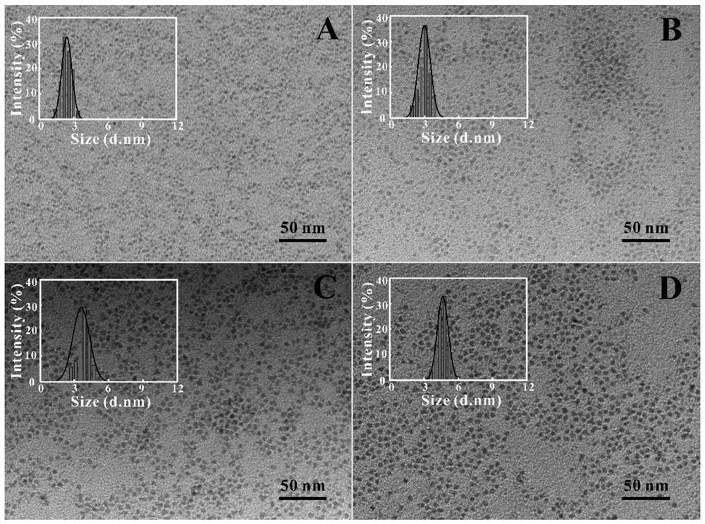
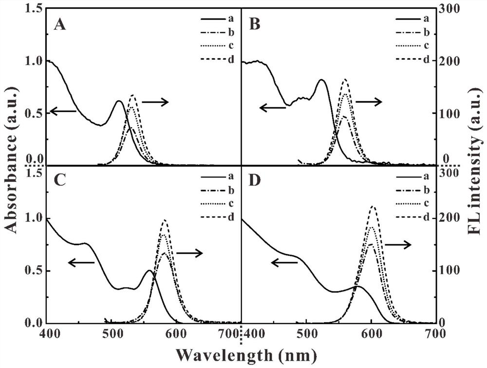
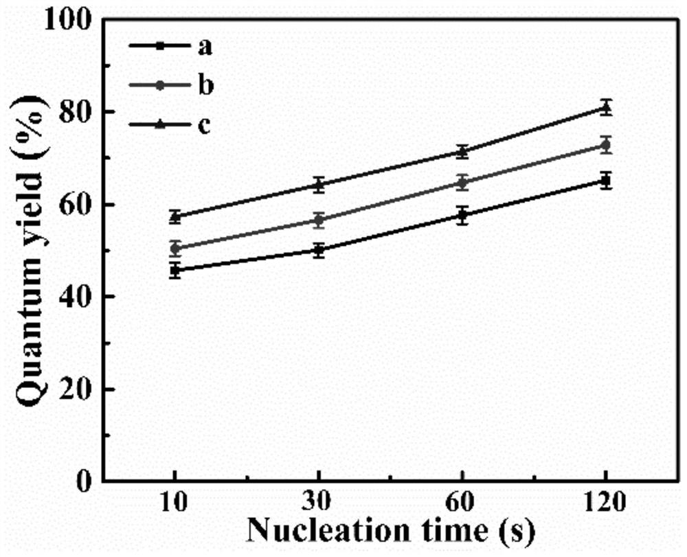
[0033] Add 0.125g of Cd(OAc) to a four-neck flask filled with argon 2 , 5g of HDA, 10g of tri-n-octylphosphine oxide (TOPO), then pass argon gas to vacuumize water and oxygen for 5 minutes each, repeat this process three times, and then pass high-purity argon gas for 10 minutes (the purity of argon gas is not less than 99.99% %), then the mixture was heated to 260 °C, stirred for 2 h and then rose to 340 °C, then dropped to 330 °C. The Se precursor solution (0.043g of Se powder dissolved in 2mL of trioctylphosphine (TOP)) was quickly injected into the four-necked flask at one time. After a certain period of nucleation, the reaction heating device was immediately removed, and the pre-cooled 20mL of toluene was used to cool the reaction solution below 140°C. After cooling to room temperature, an equal volume of methanol was added to precipitate and centrifuge (10000r...

Embodiment 2

[0056] 1. Preparation of CdTe quantum dots

[0057] Preparation of NaHTe

[0058] Weigh 51.6mg Te powder and 74.6mg NaBH 4 In the vial, fix the rubber stopper with adhesive tape, add 2 mL of deionized water into the vial with a syringe, and then place the reaction system in a refrigerator at 4°C. During the reaction, a catheter was inserted into the reaction bottle to keep it open to the atmosphere to discharge the H produced by the reaction. 2 . After about 8 hours, the black Te powder disappeared, and a white precipitate appeared at the bottom of the reaction bottle. The solution was light purple.

[0059] Synthesis of TGA-CdTe

[0060] 100mL deionized water Ar deoxygenation 30min, add 92mg CdCl 2 2.5H 2 O, 0.07mL TGA, magnetic stirring to fully dissolve. Adjust the pH to 11 using 1 mol / L NaOH solution. Add 1 mL of the above NaHTe solution. Reflow at 100°C is sufficient. The solution was taken at 1,2,3 and 4h respectively.

[0061] The CdTe quantum dots synthesiz...

Embodiment 3

[0072] 1. Synthesis of carbon quantum

[0073] Weigh 3g of citric acid and 2g of urea in 15mL of deionized water, magnetically stir to dissolve, transfer to a 25mL autoclave, heat at 180°C for 10h, cool it down to room temperature after the reaction, centrifuge the reactant at 10,000rpm for 20min, discard the precipitate, The collected supernatant was dialyzed with a dialysis bag with a molecular weight of 1000, and dried in a freeze dryer to obtain blue-emitting carbon quantum dots (CQDs).

[0074] The carbon quantum dots synthesized by the hydrothermal method are similar to spherical, with narrow particle size distribution and no obvious agglomeration. Carbon quantum dots have obvious excitation wavelength dependence, their excitation spectrum is broad and continuous, and their emission spectrum is narrow and symmetrical.

[0075] 2. Synthesis of carbon quantum dot fluorescent probes

[0076] Take 30 μL of carbon quantum dot solution, add 20 μL of EDC solution (60 mg / mL) a...


PUM


Abstract

The invention belongs to the field of biological imaging of nanomaterials, and discloses a nano fluorescent probe solution for liver cell labeling below freezing point and its application. The nano fluorescent probe solution includes nano fluorescent probes dispersed therein as a dispersoid; The needle includes nano fluorescent particles and targeting molecules coupled on the surface of the nano fluorescent particles, the targeting molecules can be used to target specific cells in the liver; and the nano fluorescent probe solution also includes antifreeze substances, the concentration of the antifreeze substances satisfies the nano Fluorescent probe solutions remain liquid at temperatures below freezing. In the present invention, by improving the detailed structure and composition of the nano fluorescent probe solution, the specific liver cells of the pre-selected cell type can be used as the target cells, and based on the nano fluorescent probe, the liver cells can be immunofluorescently labeled below freezing point , has the characteristics of rapidity, simplicity and high efficiency, and is especially suitable for use as a fluorescent marker in liver micro-optical section tomography.

Description

technical field [0001] The invention belongs to the field of biological imaging of nanomaterials, and more specifically relates to a nano fluorescent probe solution for labeling liver cells below freezing point and its application. Background technique [0002] Due to the advantages of strong specificity, high sensitivity, and fast detection speed, immunofluorescence technology can accurately locate cells, so it can be used in micro-optical section tomography of the liver. In immunofluorescence histochemistry studies, it is often necessary to prepare tissue sections for immunofluorescence staining to visualize cells. Frozen sections have been adopted by many researchers due to their advantages of simple operation, time-saving and fast, less environmental pollution, less antigenic immunogenic damage, and complete RNA information. Using immunofluorescence technology to perform fluorescent labeling and imaging of frozen sections is an important aspect in immunohistochemical res...

Claims


Application Information

Patent Timeline
Patent Type & Authority Patents(China)
IPC IPC(8): G01N33/53C09K11/02C09K11/88B82Y20/00B82Y30/00B82Y40/00G01N33/569G01N33/58G01N21/64
CPCC09K11/02C09K11/883B82Y20/00B82Y30/00B82Y40/00G01N33/588G01N33/585G01N33/582G01N33/56966G01N21/64
Inventor 赵元弟何艳飞刘波
Owner HUAZHONG UNIV OF SCI & TECH
Why Eureka
Try Eureka