
Acid phosphatase optical fiber biosensor as well as preparation method and application thereof

A technology of acid phosphatase and biosensor, applied in instruments, optics, light guides, etc., can solve problems such as false positives and increased refractive index of the surrounding environment of optical fibers

Active Publication Date: 2021-07-30
JINAN UNIVERSITY
View PDF9 Cites 0 Cited by
  • Summary
  • Abstract
  • Description
  • Claims
  • Application Information

AI Technical Summary

Problems solved by technology

Therefore, in practical applications, it is often difficult to judge whether the target measured object or the interference object causes the increase in the refractive index of the surrounding environment of the optical fiber, which may cause a "false positive" diagnosis

Method used


Image

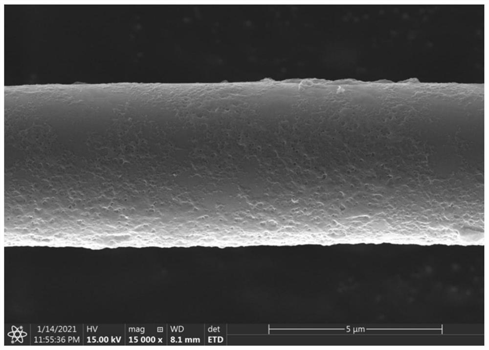
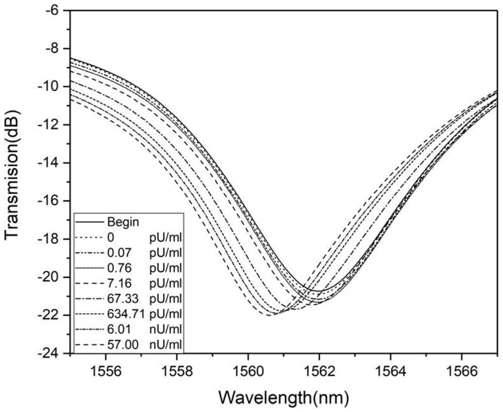
  • Acid phosphatase optical fiber biosensor as well as preparation method and application thereof
  • Acid phosphatase optical fiber biosensor as well as preparation method and application thereof
  • Acid phosphatase optical fiber biosensor as well as preparation method and application thereof

Examples


Embodiment 1

[0041] S1. Immerse the micro-nano optical fiber interferometer in the piranha solution for 60 minutes to activate the hydroxyl groups on the surface of the optical fiber. Wherein the preparation method of the piranha solution is as follows: take 7 milliliters of concentrated sulfuric acid in a beaker, slowly drop into 3 milliliters of hydrogen peroxide, and stir evenly.

[0042] S2. Dissolve 10 mg of chitosan in 20 ml of sodium acetate buffer (0.05M, pH 5.0) to prepare a 0.5 mg / ml chitosan solution. Similarly, dissolve 10 mg of sodium tripolyphosphate in 20 ml of sodium acetate buffer (0.05M, pH 5.0) to prepare a 0.5 mg / ml sodium tripolyphosphate solution. 0.5mg / ml chitosan solution and 0.5mg / ml sodium tripolyphosphate solution were mixed at a volume ratio of 1:1 to form a suspension.

[0043]S3. Immerse the micro-nano fiber interferometer with activated hydroxyl groups on the surface into the suspension in step S2 for 30 minutes, and modify the surface of the micro-nano fibe...


PUM


Abstract

The invention belongs to the technical field of biological detection, and particularly discloses an acid phosphatase optical fiber biosensor as well as a preparation method and application thereof. The preparation method of the acid phosphatase optical fiber biosensor comprises the following steps: (1) soaking a micro-nano optical fiber interferometer in a piranha solution, and activating hydroxyl on the surface of the micro-nano optical fiber interferometer; (2) mixing a solution containing an acid phosphatase substrate and a cationic polyelectrolyte solution to form an ionic cross-linked compound suspension; and (3) soaking the micro-nano optical fiber interferometer with activated hydroxyl on the surface in the suspension, and forming an acid phosphatase sensitive film specifically hydrolyzed by acid phosphatase on the surface of the micro-nano optical fiber to obtain the acid phosphatase optical fiber biosensor. Compared with a traditional electrochemical sensor, the optical fiber biosensor has the advantages of electromagnetic interference resistance and corrosion resistance, and can be applied to acid phosphatase in-vivo real-time monitoring. The detection process is not only label-free for the detection sample, but also has the advantages of simplicity, convenience, rapidness and the like.

Description

technical field [0001] The invention belongs to the technical field of biological detection, in particular to an acid phosphatase optical fiber biosensor, a preparation method and an application. Background technique [0002] Acid phosphatase is an enzyme that can hydrolyze phosphate molecules under acidic conditions to release free phosphate groups. In the human body, acid phosphatase is produced by tissues such as the prostate, liver, spleen, and bone marrow, and is widely present in various tissues, cells, and body fluids. Acid phosphatase is usually present in low concentrations in the human body, however, in some diseases, acid phosphatase synthesis is significantly altered, and its over- or under-expression of the enzyme is considered part of the pathological process. Abnormal levels of acid phosphatase can be used as an indication of lysosomal acid phosphatase deficiency, giant cell tumor of bone, hemolytic anemia, childhood developmental disorders, Gaucher's disease...

Claims


Application Information

Patent Timeline
IPC IPC(8): G01N21/45G02B6/255
CPCG01N21/45G02B6/255G01N2021/458
Inventor 王维李泽森洪美静蔡洁旋关柏鸥
Owner JINAN UNIVERSITY
Learn More