Tumor marker screening method based on nucleosome distribution characteristics and application

A technology of tumor markers and screening methods, applied in the field of quantification of nucleosome distribution, can solve problems such as screening of tumor biomarkers

Pending Publication Date: 2021-09-07
BERRY ONCOLOGY CO LTD +1
View PDF0 Cites 2 Cited by
  • Summary
  • Abstract
  • Description
  • Claims
  • Application Information

AI Technical Summary

Problems solved by technology

However, this study can only reflect that nucleosome protection has a certain potential in speculatively expressed genes, and cannot solve the problem of tumor screening, especially the screening of early tumors and the screening of related biomarkers

Method used


Image

  • Tumor marker screening method based on nucleosome distribution characteristics and application
  • Tumor marker screening method based on nucleosome distribution characteristics and application
  • Tumor marker screening method based on nucleosome distribution characteristics and application

Examples


Embodiment 1

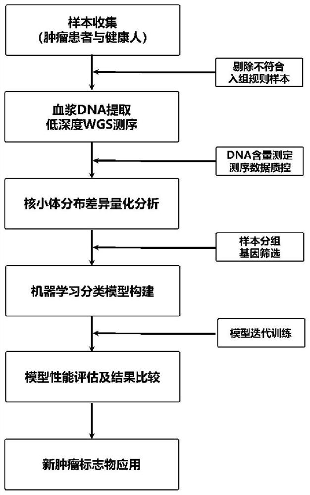
[0096] Determine the enrolled tumor patients and healthy people. A total of 984 subjects were recruited from 508 patients with liver cancer (HCC) and 476 healthy controls (NC) from two centers. According to the pathological diagnosis, 27 liver cancer samples were excluded, and 957 subjects were finally determined ( figure 1 ).

[0097] Plasma cell-free DNA (cfDNA) was extracted from tumor patients and healthy people. Take 3ml of peripheral blood from each participant (collected and stored in Streck cell-free DNA blood collection tubes), use Eppendorf centrifuges (5810R and 5427R, German), and centrifuge at 1600g at low speed for 10min at a low temperature of 4°C, and only take the supernatant liquid; then centrifuged at 16000 g for 10 min at high speed, and the supernatant was taken to obtain a plasma sample. The free DNA in the plasma was extracted with a kit MagMAX Cell-Free DNA Isolation Kit (Thermo) and a nucleic acid extractor (Thermo Kingfisher FLEX, USA). The concen...

Embodiment 2

[0099] Low-depth whole-genome sequencing was performed on the cfDNA samples of all participants prepared in Example 1. The sequencing process is as follows:

[0100] (1) WGS library construction and on-machine sequencing: take 5ng cfDNA and use the Enzymatics (USA) kit to construct a pre-library, mainly including two steps of end repair (5X ER / A-Tailing Enzyme Mix) and adapter (WGS Ligase) , the adapter sequence is suitable for the Illumina NovaSeq 6000 sequencing platform. The adapters were ligated and purified using XP magnetic beads (Agencourt AMPure XP beads, Beckman Coulter). The concentration value of WGS library was determined by qPCR (KAPA Library QuantKit, Roche), and the library size was determined by Fragment Analyzer (Agilent, USA). Afterwards, paired-end 150bp sequencing was performed on the Illumina NovaSeq 6000 sequencing platform, and the average data volume of a single sample was 2X that of the whole genome.

[0101] (2) Data quality control: Use Fastp soft...

Embodiment 3

[0103] The sequencing data obtained in Example 2 was used to quantify the difference in nucleosome distribution in the transcription start site region of the genome-wide gene, and the specific steps were as follows:

[0104] (1) Acquisition of the gene transcription start site region of the whole genome: use the transcription start site of the main transcript of the reference gene published in the USCS database, and extend 2500 bp before and after each as the transcription start site region of the gene.

[0105] (2) Acquisition of the sequencing depth of the transcription start site region: divide the transcription start site region into 500 small regions, and use the average sequencing depth of each small region to represent the sequencing depth of the small region, thereby eliminating the sequencing depth Insufficient results in some sites not being covered by DNA fragments.

[0106](3) Delineation of the central area of ​​the transcription start site, the edge area and the ...


PUM


Abstract

The invention discloses a screening method of a tumor marker or a tumor marker combination. The screening method comprises the following steps: i) determining the distribution condition of nucleosomes; and / or ii) screening a tumor-related marker through one or more indexes including the nucleosome distribution condition. The invention also provides a tumor screening model construction method based on the screening method, a cancer screening, risk prediction and / or diagnosis method, and a related tumor marker combination, a kit, a system, a device, a computer readable storage medium and equipment.

Description

technical field [0001] The invention belongs to the technical field of gene detection, and in particular relates to a tumor marker screening method based on nucleosome distribution characteristics and a related tumor screening model construction method, cancer screening, risk prediction and / or diagnosis method, system, and device , a computer-readable storage medium, a device, and a related method for quantifying nucleosome distribution. Background technique [0002] Cancer is one of the three major diseases that seriously endanger human health in the world. According to reports, there were 18.19 million new cancer cases and 9.6 million cancer deaths in the world in 2018, and the cancer prevalence rate in my country is at the upper-middle level in the world. WHO believes that one third of cancers can be cured with early detection. Compared with late-stage cancers, early-stage cancers have not metastasized and are easier to remove through surgery, radiotherapy, chemotherapy,...

Claims


Application Information

Patent Timeline
IPC IPC(8): G16B30/00G16B20/50G16B20/20G16B20/10G16H50/20
CPCG16B30/00G16B20/50G16B20/10G16B20/20G16H50/20
Inventor 张清政白健吴琳
Owner BERRY ONCOLOGY CO LTD
Why Eureka
Try Eureka