Electron energy loss spectroscopy analysis method for characterization of fine structure of transition metal oxide

A technology of electron energy loss and transition metals, applied in material analysis using radiation diffraction, material analysis using radiation, etc., can solve problems such as low K-shell signal, restricted coordination structure, complex electronic structure of L-shell, etc. , to achieve high spatial resolution, high sensitivity, and cost-effective effects

Active Publication Date: 2021-09-17
INST OF METAL RESEARCH - CHINESE ACAD OF SCI
View PDF9 Cites 1 Cited by
  • Summary
  • Abstract
  • Description
  • Claims
  • Application Information

AI Technical Summary

Problems solved by technology

However, due to the low K-shell signal and the complex electronic structure of the L-shell of metal elements in transition metal oxides, it is limited to obtain information on the coordination structure through the electron energy loss spectrum of metal elements.

Method used


Image

  • Electron energy loss spectroscopy analysis method for characterization of fine structure of transition metal oxide
  • Electron energy loss spectroscopy analysis method for characterization of fine structure of transition metal oxide
  • Electron energy loss spectroscopy analysis method for characterization of fine structure of transition metal oxide

Examples


Embodiment 1

[0027] This embodiment is used for the electron energy loss spectroscopy analysis method of commercial FeO microstructure characterization, and the specific process is as follows:

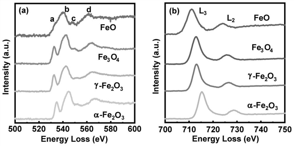
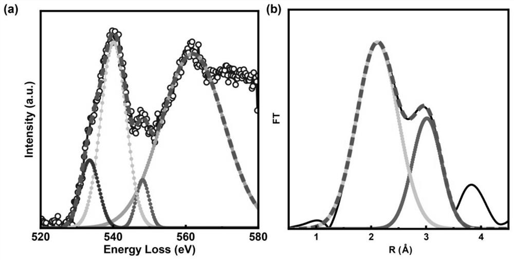
[0028] 1. Through the FEI Tecnai G equipped with a Gatan Imaging Filter (GIF) 965 spectrometer 2 The electron energy loss spectrum of the oxygen of commercial FeO was collected in the F20 TEM (see figure 1). Then Gaussian fitting was performed on the four peaks in FeO (approximately in the range of 530eV-580eV) by DigitalMicrograph software (see figure 2 ). The valence state of Fe can be calculated as +2.00 through the peak intensity ratio of the front peak (530-540eV) and the highest peak (540-550eV).

[0029] 2. Obtain the oxygen atom coordination structure through the extended fine structure of the electron energy loss spectrum of FeO: it is necessary to extract the oscillation signal in the extended fine structure of the electron energy loss spectrum of oxygen, and then perform signal denoi...

Embodiment 2

[0033] This embodiment is used for Fe 3 o 4 Electron energy loss spectroscopy analysis method for nanorod microstructure characterization, the specific process is as follows:

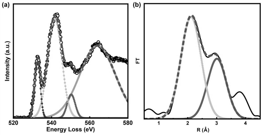
[0034] 1. Through the FEI Tecnai G equipped with a Gatan Imaging Filter (GIF) 965 spectrometer 2 Fe was collected in the F20 transmission electron microscope 3 o 4 The electron energy loss spectrum of the oxygen of the nanorods (see figure 1 ). Then by DigitalMicrograph software for Fe 3 o 4 The four peaks in the nanorods (approximately in the range of 530eV to 580eV) were Gaussian fitted (see image 3 ). The average valence state of Fe can be calculated by the peak intensity ratio of the front peak (530-540eV) and the highest peak (540-550eV) to be +2.69.

[0035] Second, through Fe 3 o 4 The oxygen electron energy loss spectrum extended fine structure of nanorods is obtained to obtain the oxygen atom coordination structure: it is necessary to extract the oscillation signal in the oxygen ele...

Embodiment 3

[0039] This embodiment is used for γ-Fe 2 o 3 Electron energy loss spectroscopy analysis method for nanorod microstructure characterization, the specific process is as follows:

[0040] 1. Through the FEI Tecnai G equipped with a Gatan Imaging Filter (GIF) 965 spectrometer 2 γ-Fe was collected in F20 transmission electron microscope 2 o 3 The electron energy loss spectrum of the oxygen of the nanorods (see figure 1 ). Then through DigitalMicrograph software for γ-Fe 2 o 3 The four peaks in the nanorods (approximately in the range of 530eV to 580eV) were Gaussian fitted (see Figure 4 ). The valence state of Fe can be calculated as +3.04 through the peak intensity ratio of the front peak (530-540eV) and the highest peak (540-550eV).

[0041] 2. Through γ-Fe 2 o 3 The oxygen electron energy loss spectrum extended fine structure of nanorods is obtained to obtain the oxygen atom coordination structure: it is necessary to extract the oscillation signal in the oxygen elec...


PUM


Abstract

The invention discloses an electron energy loss spectroscopy analysis method for transition metal oxide microstructure characterization, and belongs to the technical field of electron microscopic analysis. The method comprises the following steps: firstly, acquiring an electron energy loss spectrum of oxygen in a transition metal oxide by using a transmission electron microscope, and then carrying out Gaussian function fitting on a near-edge fine structure of the electron energy loss spectrum of oxygen to obtain valence information of metal elements in the material; then, by means of quintic spline function fitting, extracting an oscillation signal in the oxygen electron energy loss spectrum extension fine structure, and carrying out coordinate transformation, denoising and amplitude modulation processing on the oscillation signal; and then carrying out Fourier transform on the processed oscillation signal to obtain oxygen atom coordination structure information. combining metal element valence state information and oxygen atom coordination structure information to finally obtain complete quantitative information about the transition metal oxide microstructure. The analysis method is helpful for research of transition metal oxide materials in the fields of catalysis and energy storage.

Description

technical field [0001] The invention relates to the technical field of electron microscopy analysis, in particular to an electron energy loss spectroscopy analysis method for characterization of the fine structure of transition metal oxides. Background technique [0002] Transition metal oxides have attracted extensive attention as catalysts or electrode materials due to their advantages of multiple valence states and multiphase transitions. In order to explore the reaction kinetics of such materials in catalytic and electrode processes, it is necessary to analyze the changes in the microstructure of transition metal oxides. In general, XRD and in-situ XRD are used to obtain crystal phase transition information of materials. Using X-ray photoelectron spectroscopy (XPS) and X-ray absorption fine structure (XAFS), the valence state evolution and coordination environment of the characteristic elements of transition metal oxides can be obtained. However, none of these methods ...

Claims


Application Information

Patent Timeline
Patent Type & Authority Applications(China)
IPC IPC(8): G01N23/04G01N23/20
CPCG01N23/04G01N23/20
Inventor 张炳森陈隽楠
Owner INST OF METAL RESEARCH - CHINESE ACAD OF SCI
Why Eureka
Try Eureka