
Flexible high-elasticity auricular cartilage scaffold capable of accurately reproducing cranium-ear angle

A technology of cartilage and elastic ears, which is applied in ear treatment, ear implants, 3D object support structures, etc. It can solve the problems of infection, large pores of the bracket, etc., and achieve the goal of raising the auricle, real hand feeling, and shortening the operation cycle. Effect

Inactive Publication Date: 2021-09-21
必优提苏州数字医疗科技有限责任公司
View PDF3 Cites 0 Cited by
  • Summary
  • Abstract
  • Description
  • Claims
  • Application Information

AI Technical Summary

Problems solved by technology

However, the costal cartilage scaffolds currently used by most patients have large pores in the scaffold, and a small amount of bleeding is not easy to be found. If it is not treated in time, there will be a risk of infection

Method used


Image

  • Flexible high-elasticity auricular cartilage scaffold capable of accurately reproducing cranium-ear angle
  • Flexible high-elasticity auricular cartilage scaffold capable of accurately reproducing cranium-ear angle

Examples


Embodiment Construction

[0033] The present invention will be further described below through specific embodiments.

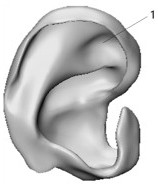
[0034] (1) Obtain CT slice images of the reference auricle shape. In this case, the left auricle of a 7-year-old boy is deformed and the right is deformed.

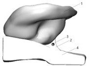
[0035] (2) Perform image processing on the obtained slice image, extract the outline slice image of the ear cartilage, and obtain the three-dimensional solid model of the ear cartilage after three-dimensional reconstruction. The software used in this example is Mimics21.0 from Materialise Company in Belgium.

[0036] (3) Turn on the Threshold function, the soft tissue threshold range is between -680 and 110, and generate a corresponding mask.

[0037] (4) Use the Crop Mask to cut out the auricle of the patient's healthy ear and the part of the skin connected to the temporal bone, and manually erase the noise points.

[0038] (5) Using the Dynamic Region Growth function, because the threshold range of ear cartilage is between 50-...


PUM


Abstract

The invention relates to design and rapid preparation of an implantable auricular cartilage scaffold. Existing methods for preparing auricular cartilage scaffolds have the defects of large trauma, hard hand feeling, poor recovery effect and the like. The method for designing and preparing the auricular cartilage scaffold comprises the following steps: 1, obtaining a two-dimensional lamellar image of a reference auricle; 2, obtaining an auricular cartilage three-dimensional solid model; 3, adding a supporting bracket to the ear back of the bracket according to the cranium-ear angle of the healthy ear of the patient; 4, designing an inner flow channel in the supporting structure; and 5, inputting the designed three-dimensional solid model into additive manufacturing equipment in layers, and carrying out layer-by-layer accumulation by the equipment to obtain the auricular cartilage scaffold. The auricular cartilage scaffold prepared by the method can be implanted into a body, and is vivid in hand feeling, and the appearance of the auricular cartilage stent is symmetrical to that of a healthy auricle.

Description

technical field [0001] The invention is a flexible and high-elastic ear cartilage support for accurately reproducing cranial and ear angles, and belongs to the fields of medical materials, plastic surgery and rapid prototyping. Background technique [0002] Microtia is a common congenital malformation with an incidence rate of 5 / 10000 and a male to female ratio of 2:1. This disease has seriously affected the physical and mental health of patients and their families. Congenital microtia is treated by auricle reconstruction surgery. Due to the complex three-dimensional structure of the outer ear, it is one of the most complicated and difficult operations in plastic surgery. [0003] At present, the main surgical method for treating microtia is the skin expansion method, and the material used is the patient's costal cartilage. The operation can be divided into three phases: in the first phase, the skin dilator is implanted in the mastoid area behind the affected side to form ...

Claims


Application Information

Patent Timeline
Patent Type & Authority Applications(China)
IPC IPC(8): A61F2/18A61F2/30A61F11/00B29C64/118B29C64/386B29C64/393B33Y10/00B33Y40/00B33Y50/00B33Y50/02
CPCA61F2/18A61F2/30756A61F2/30942B29C64/118B29C64/386B29C64/393B33Y10/00B33Y40/00B33Y50/00B33Y50/02A61F2002/183A61F2002/30948A61F2002/30985A61F2002/30943
Inventor 史廷春闫毅文岳秀艳
Owner 必优提苏州数字医疗科技有限责任公司
Learn More