High-lysis klebsiella pneumoniae bacteriophage RDP-KP-20007 and application thereof
A technology of RDP-KP-20007, Klebsiella pneumoniae bacteriophage, applied in bacteriophage, virus/phage, medical raw materials derived from virus/phage, etc. properties, etc., to achieve good application prospects and strong cleavage activity.
- Summary
- Abstract
- Description
- Claims
- Application Information
AI Technical Summary
Problems solved by technology
Method used
Image
Examples
Embodiment 1
[0033] Example 1 Isolation and identification of pathogenic Klebsiella pneumoniae BK-20007
[0034]Samples were taken from livers of chickens with Klebsiella pneumoniae disease, inoculated in DHL medium by aseptic operation, and cultured at 37°C for 18-24h. Klebsiella pneumoniae forms pale pink colonies on the DHL agar plate, with a large raised surface, smooth and moist, in the form of mucus. Adjacent colonies are easy to fuse into a thick juice, and long filaments can be pulled out when the inoculation needle is picked.
[0035] Select the pale pink colonies to streak and purify three times in a row for biochemical identification. Transplanted from DHL medium to Klebsiella medium and cultured at 37°C for 18-24h, the slant surface produced acid, the bottom layer produced acid and gas, and the hydrogen sulfide was negative. Biochemical identification showed positive V-P test, positive nitrate reduction test, negative M.R, positive Simon's citrate utilization test, positive ma...
Embodiment 2
[0037] Example 2 Isolation and identification of Klebsiella pneumoniae phage RKP-KP-20007
[0038] The liver sample used in the experiment of the present invention was collected from a sick chicken in a broiler chicken farm in Jimo, Qingdao in 2020, and used as a sample for phage isolation.
[0039] (1) Isolation of bacteriophage RKP-KP-20007:
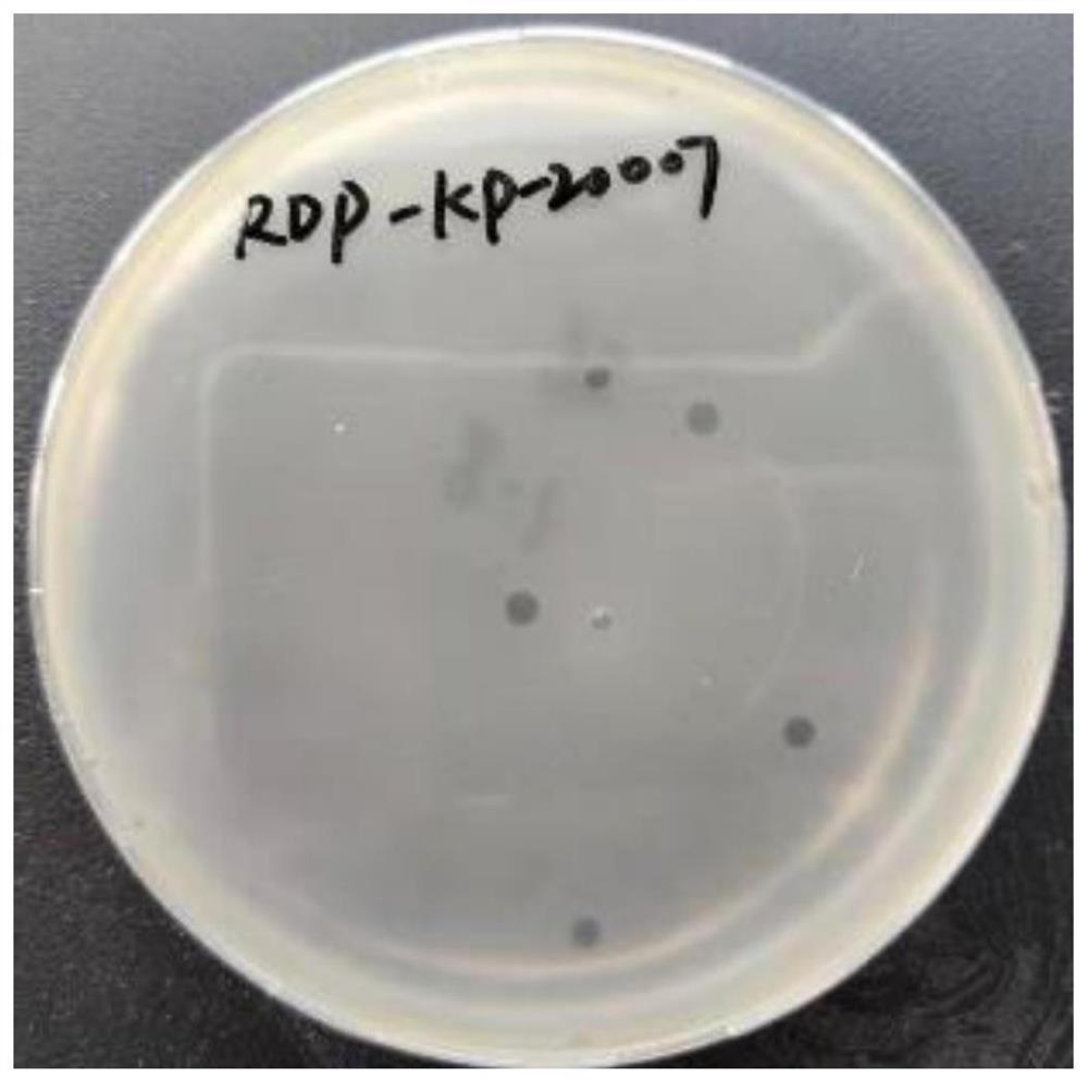
[0040] Take 10㎎ of the liver and soak it in 10ml LB broth overnight at 4°C, centrifuge at 10,000rpm for 5min, and filter the supernatant with a 0.22μm microporous membrane to sterilize. Take 3ml of the filtrate and 3ml of the host bacterium culture solution in the logarithmic growth phase, add them together to 20ml of autoclaved LB broth, put them in a 37°C incubator, and culture them overnight to multiply the possible phages. Take 3ml of the sample culture solution, centrifuge at 10000rpm for 5min, and take the supernatant to filter and sterilize with a 0.22μm microporous membrane. Assume that the above filtrate contains phages. Th...
Embodiment 3
[0046] Morphological observation of embodiment 3 phage
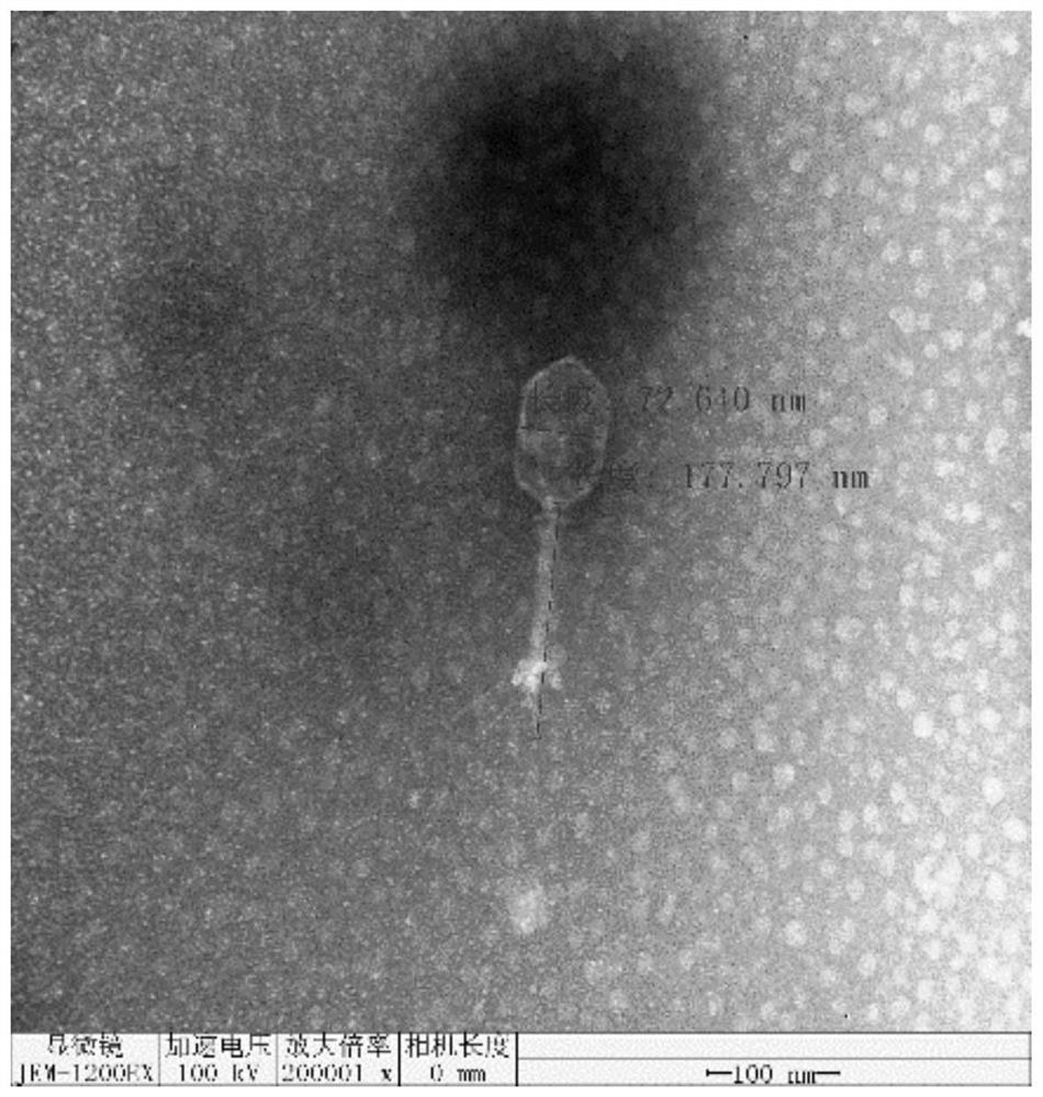
[0047] Using phosphotungstic acid negative staining method: 20 μL of phage proliferation solution (titer 10 11 pfu / mL) was dropped on a paraffin sheet, and the copper grid was placed on it for 10 minutes and then removed to absorb phage. Dry at natural room temperature for 2-3 minutes, stain with 2% phosphotungstic acid aqueous solution, blot the water after 2 minutes, dry naturally for 5 minutes, and observe the phage morphology with an electron microscope.
[0048] Photo by electron microscope ( figure 2 ) It can be seen that the head of bacteriophage RKP-KP-20007 is icosahedral and stereosymmetric, the length of the head is 72.640nm, and the length of the phage tail is 177.797nm. .
PUM
 Abstract
Description
Claims
Application Information
 - R&D
- Intellectual Property
- Life Sciences
- Materials
- Tech Scout
- Unparalleled Data Quality
- Higher Quality Content
- 60% Fewer Hallucinations
Browse by: Latest US Patents, China's latest patents, Technical Efficacy Thesaurus, Application Domain, Technology Topic, Popular Technical Reports.
© 2025 PatSnap. All rights reserved.Legal|Privacy policy|Modern Slavery Act Transparency Statement|Sitemap|About US| Contact US: help@patsnap.com


