Cancer cell xenograft zebrafish model and construction method and application thereof
A technology of xenotransplantation and construction method, which is applied in the field of cancer cell xenotransplantation zebrafish model, can solve problems such as judging the proliferation of cancer cells, and achieve the effects of reducing damage, ensuring normal survival, and improving survival
- Summary
- Abstract
- Description
- Claims
- Application Information
AI Technical Summary
Problems solved by technology
Method used
Image

Examples
Embodiment 1
[0028] Example 1: Obtaining fluorescently labeled cancer cells
[0029] 1) Cell recovery: Take out a tube of frozen liver cancer 468 cells from the -80°C refrigerator, quickly place them in a 37°C water bath, place the thawed liver cancer 468 cells in a centrifuge tube, add 1ml of cell culture medium, and centrifuge , aspirate the supernatant and continue to add cell culture medium, fully resuspend, transfer to a culture dish, make up 7ml of cell culture medium, and culture in a cell culture incubator;
[0030] 2) Cell subculture: When the cells have covered about 70-80% of the bottom of the culture dish, first suck off the cell culture medium, wash the cells 2-3 times with 2ml 1×PBS, and cover the bottom with 0.25% trypsin Put it in the cell culture incubator for 3-4 minutes, take out the cell culture medium, then transfer it to a centrifuge tube, centrifuge, suck out the supernatant and continue to add the cell culture medium, fully resuspend, and culture in the cell culture...
Embodiment 2
[0032] Example 2: Construction of cancer cell xenograft zebrafish model
[0033] 1) Cell counting: Take a small amount of cells to count before injection, resuspend the labeled liver cancer 468 cells in 2 mL 1×PBS, take out 10 μL of the suspension and add 90 μL of 1×PBS to dilute 10 times, mix well, and inhale 10 μL of the diluted suspension Gently blow the solution into the cell counting plate and count under the microscope: (33+40+36+25) / 4×10×10 4 =3.35×10 6 / mL, the average cell density of the cell suspension is 3.35×10 6 / mL.
[0034]2) Injection: Collect fertilized eggs of the AB strain, wait for the embryos to grow to 48hpf (hours after fertilization), anesthetize with tricaine, and place them on a gel plate made of agarose. Manually peel off the film. Adjust the cell culture medium for the fluorescently labeled cells in Example 1 to 2~3×10 4 / μL density. Prepare a microinjection capillary needle for microinjection into the pericardial space of 48hpf zebrafish embr...
Embodiment 3
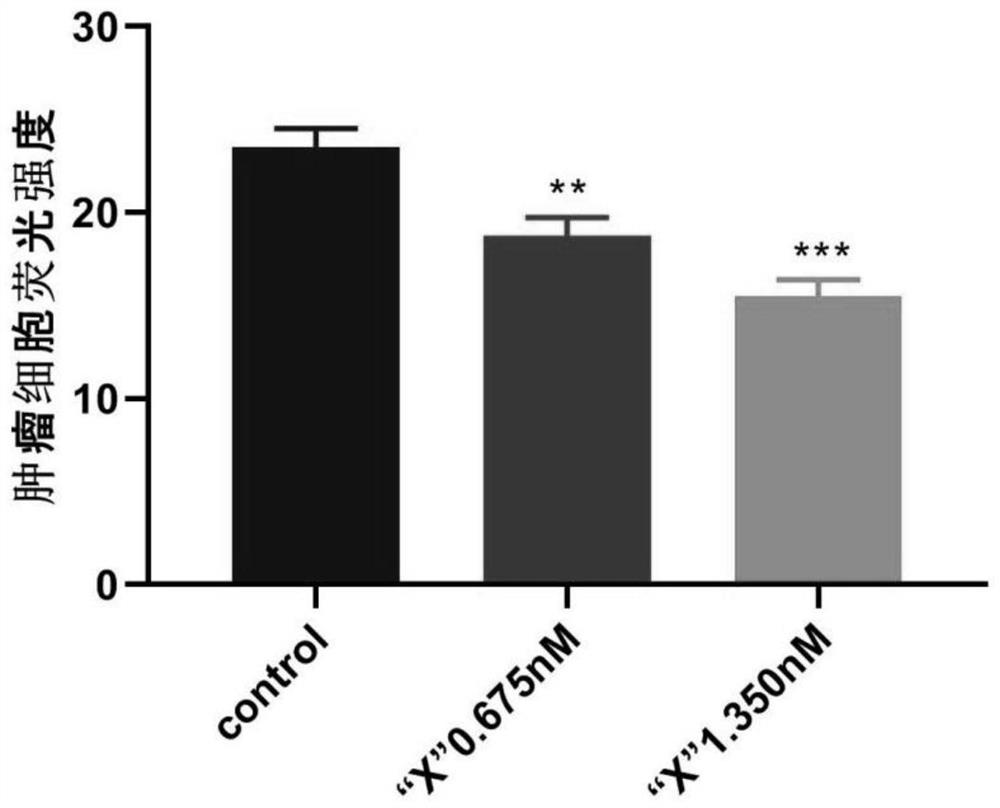
[0036] Drug treatment after screening: Observe the injected embryos carrying green fluorescence under a fluorescent microscope, select embryos with normal embryonic development and good fluorescence status, and randomly divide them into three groups. Wherein two groups adopt the drug treatment of 0.675nM and 1.350nM concentration respectively, remaining a group is as blank control group (control), and drug treatment 24h and 48h are sampled respectively. The medicine is represented by X, and Apatinib is selected to carry out drug treatment to the zebrafish in the embodiment of this case.
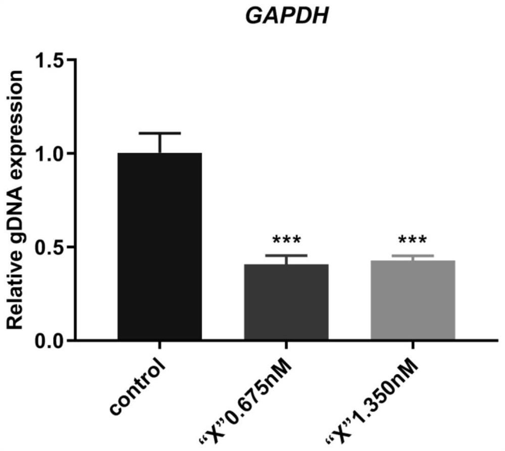
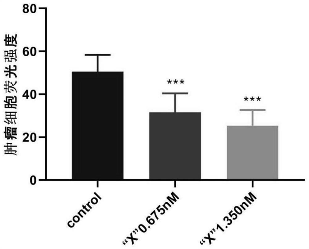
[0037] 1. Directly observe the inhibitory effect of drugs on tumors through fluorescence microscopy, such as figure 2 and image 3 Schematic diagrams of the fluorescence expression of liver cancer 468 cells in zebrafish after 24 hours and 48 hours of drug treatment, respectively. The fluorescence expression of liver cancer 468 cells with fluorescent labels in the pericardial space of zebraf...
PUM
 Abstract
Description
Claims
Application Information
 - R&D
- Intellectual Property
- Life Sciences
- Materials
- Tech Scout
- Unparalleled Data Quality
- Higher Quality Content
- 60% Fewer Hallucinations
Browse by: Latest US Patents, China's latest patents, Technical Efficacy Thesaurus, Application Domain, Technology Topic, Popular Technical Reports.
© 2025 PatSnap. All rights reserved.Legal|Privacy policy|Modern Slavery Act Transparency Statement|Sitemap|About US| Contact US: help@patsnap.com