Apoptotic vesicle as well as preparation method and application thereof
A technology of microvesicles and apoptosis, applied in biochemical equipment and methods, cell dissociation methods, microorganisms, etc., can solve problems such as limiting therapeutic applications and side effects, and achieve simple extraction methods, improved adipogenic ability, and yield high effect
- Summary
- Abstract
- Description
- Claims
- Application Information
AI Technical Summary
Problems solved by technology
Method used
Image
Examples
Embodiment 1
[0032] Example 1 Efficient extraction of apoVs derived from RAW264.7
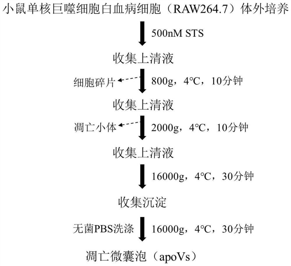
[0033] RAW264.7 was cultured in vitro, and when the cell confluence reached 90-100%, 500nM STS was added to induce apoptosis, and the apoVs derived from RAW264.7 were obtained by gradient centrifugation, and its concentration was detected by nanoparticle tracking analysis, and its protein was detected by BCA method Quantity, obtain the optimal extraction conditions, and establish a standard extraction process. details as follows:
[0034] a) Centrifuge the cell culture supernatant at 800 g for 10 min at 4° C. to remove cell debris in the culture supernatant, and take the supernatant to obtain the first centrifugation supernatant;
[0035] b) Centrifuge the first centrifugation supernatant at 4° C. and 2000 g for 10 min to remove impurities such as apoptotic bodies in the first centrifugation supernatant, and take the supernatant to obtain the second centrifugation supernatant ;
[0036] c) centrifuging t...
Embodiment 2
[0038] Example 2 Characteristic analysis of apoVs derived from RAW264.7
[0039] The morphology, particle size, and concentration of apoVs derived from RAW264.7 were detected by cryo-TEM and nanoparticle tracking analysis.
[0040] Cryo-TEM:
[0041] (1) Pipette 5 μl of apoptotic microvesicle suspension onto the copper grid, and let it stand at room temperature for 1 min;
[0042] (2) Use filter paper to absorb excess liquid along the outside of the copper grid, absorb 5 μl of 2% uranyl acetate and drop it onto the copper grid, and let stand at room temperature for 30 seconds;
[0043] (3) Absorb the excess liquid along the outside of the copper mesh with filter paper, and let it dry at room temperature;
[0044] (4) Images were taken under a transmission electron microscope, and the voltage was set to 120kV.
[0045] Nanoparticle size tracking analysis detection:
[0046] (1) Use a nanoparticle tracking analyzer to record the trajectory of exosomes under Brownian motion; ...
Embodiment 3
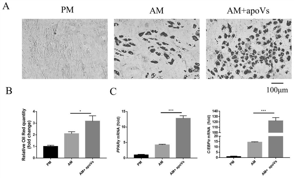
[0049] Example 3 In vitro experiments to detect the effect of apoVs derived from RAW264.7 on the adipogenic differentiation of human adipose-derived mesenchymal stem cells in vitro
[0050]After 14 days of adipogenic induction, the effect of cell adipogenic differentiation was examined by Oil Red O staining.
[0052] To prepare the dyeing solution, weigh 0.5 g of Oil Red O dry powder, dissolve it in 100 ml of 100% isopropanol, and after fully dissolved, it becomes Oil Red O stock solution, and store it in the dark at 4°C. Before staining, take Oil Red O stock solution and dilute it according to the ratio of stock solution: distilled water = 3:2. After filtering with filter paper, it becomes Oil Red O working solution, which is used for staining.
[0053] Aspirate the medium, rinse with PBS three times, and fix with 10% neutral formalin for 1 hour. Discard the formalin, rinse with PBS three times, and rinse with 60% isopropanol. After drying, add...
PUM
| Property | Measurement | Unit |
|---|---|---|
| particle diameter | aaaaa | aaaaa |
| particle size | aaaaa | aaaaa |
Abstract
Description
Claims
Application Information
 - R&D
- Intellectual Property
- Life Sciences
- Materials
- Tech Scout
- Unparalleled Data Quality
- Higher Quality Content
- 60% Fewer Hallucinations
Browse by: Latest US Patents, China's latest patents, Technical Efficacy Thesaurus, Application Domain, Technology Topic, Popular Technical Reports.
© 2025 PatSnap. All rights reserved.Legal|Privacy policy|Modern Slavery Act Transparency Statement|Sitemap|About US| Contact US: help@patsnap.com


