Device for establishing blood vessel tunnel under direct vision

A subcutaneous tunnel and blood vessel technology, applied in the field of medical devices, can solve problems such as deviation of the advancing direction, injury of iliac femoral blood vessels, infection of artificial blood vessels, etc., and achieve the effect of avoiding damage to important anatomical structures

Pending Publication Date: 2022-03-25
复旦大学附属中山医院青浦分院
View PDF8 Cites 0 Cited by
  • Summary
  • Abstract
  • Description
  • Claims
  • Application Information

AI Technical Summary

Problems solved by technology

In the past, the traditional subcutaneous vascular tunneler was used to establish a subcutaneous tunnel. This method has the following disadvantages: (1) This method establishes a vascular channel subcutaneously based on the experience of the surgeon. Since the subcutaneous advancement of the vascular tunneler cannot be directly observed, the blood vessel may be damaged. , Nerve and muscle injury, especially in the main-double-femoral bypass surgery, the advancement of the subcutaneous tunnel device may cause damage to the iliofemoral blood vessels, resulting in fatal complications
(2) If blood vessel damage leads to malabsorption of subcutaneous hematoma, it may lead to serious complications such as further infection of artificial blood vessels; (3) At the same time, due to the inability to see directly, the subcutaneous tunnel device enters different anatomical levels during the advancement process, so that after repeated pushing and subsequent The withdrawal process will aggravate the above damage (4) During the process of pushing the vascular tunnel device, because there is no visible fiber indicator at the front end, the pushing direction deviates from the position of the vascular anastomosis, so it is necessary to constantly adjust the pushing direction, which can also aggravate the above damage
However, it is about a process of directly advancing a vascular tunnel device under the skin when establishing a vascular channel under the skin, avoiding damage to blood vessels, nerves and muscles, and at the same time, it is convenient to adjust the direction of advancement, accurately align the vascular anastomosis, and avoid clinical complications. There are no relevant reports on the establishment of vascular tunneling devices under direct vision

Method used


Image

  • Device for establishing blood vessel tunnel under direct vision
  • Device for establishing blood vessel tunnel under direct vision
  • Device for establishing blood vessel tunnel under direct vision

Examples


Embodiment 1

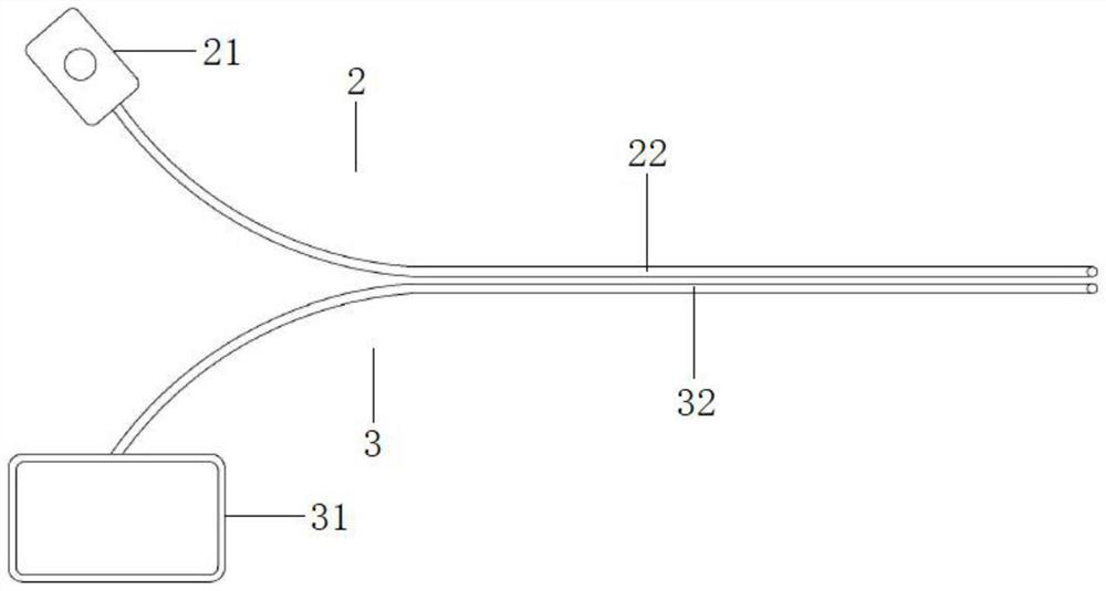
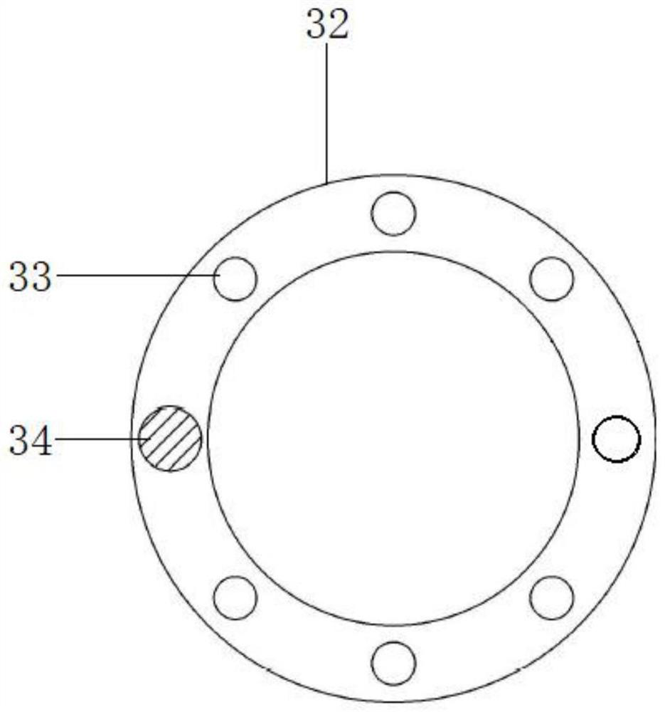
[0035] Please see attached Figure 1-3 , figure 1 It is a structural schematic diagram of a subcutaneous tunnel device for establishing a blood vessel tunnel device under direct vision according to the present invention, figure 2 It is a structural schematic diagram of an inflatable device and an electron microscope for establishing a vascular tunnel device under direct vision in the present invention, image 3 It is a schematic diagram of the structure of the front end of the fiber conduit of the device for establishing a blood vessel tunnel under direct vision of the present invention. A device for establishing a blood vessel tunnel under direct vision, the device for establishing a blood vessel tunnel under direct vision includes a subcutaneous tunnel device 1, an inflatable device 2 and an electron microscope 3; the body of the subcutaneous tunnel device 1 is a cylindrical structure, and the front end is a diameter Gradually shrinking structure; the inside of the subcut...

Embodiment 2

[0038] This embodiment is basically the same as Embodiment 1, and the difference is that a blood vessel channel (not shown) with a hollow structure is also provided in the subcutaneous tunnel device 1 in the present embodiment; 1 is also equipped with a vascular channel design, after the subcutaneous tunneler 1 is pushed to the correct position, the artificial blood vessel or autologous blood vessel can be directly successfully entered into the subcutaneous tunnel through the vascular channel without first exiting the inflatable tube 22 and the fiber tube 32, Helps to save operation time.

[0039] A device for establishing a blood vessel tunnel under direct vision of the present invention, through the design of the body of the subcutaneous tunnel device having a cylindrical structure and the front end of which the diameter gradually shrinks, can reduce the resistance of the subcutaneous tunnel device entering the subcutaneous fascia and facilitate advancement or withdrawal; By...


PUM


Abstract

The invention relates to a device for establishing a blood vessel tunnel under direct vision. The device for establishing the blood vessel tunnel under the direct vision comprises a subcutaneous tunnel device, an inflation device and an electron microscope, the subcutaneous tunnel device body is of a cylindrical structure, and the front end of the subcutaneous tunnel device body is of a structure with the diameter gradually reduced. A gas channel and a microscope channel which are of hollow structures are respectively arranged in the subcutaneous tunnel device; the inflating device comprises an inflating bag and an inflating tube; the diameter of the gas filling pipe is smaller than that of the gas channel; the electronic fiberscope comprises a display screen and a fiber pipeline; the tail end of the fiber pipeline is connected with the display screen, and an illuminating lamp and a camera are arranged in the front end of the fiber pipeline; and the diameter of the fiber pipeline is smaller than that of the microscope pipeline. The device has the advantages that when a blood vessel channel is built subcutaneously, the blood vessel tunnel device can be directly propelled subcutaneously, damage to blood vessels, nerves and muscles is avoided, meanwhile, the propelling direction can be conveniently adjusted, a blood vessel anastomotic stoma can be accurately aligned, and clinical complications are avoided.

Description

technical field [0001] The invention relates to the technical field of medical instruments, in particular to a device for establishing a blood vessel tunnel under direct vision. Background technique [0002] Lower extremity arteriosclerosis occlusive disease refers to the chronic progressive disease of intima thickening, lumen stenosis or occlusion of the lower extremity arteries due to arteriosclerosis, resulting in insufficient blood supply to the affected extremities, and is often the manifestation of systemic arteriosclerosis vascular disease in the arteries of the lower extremities . [0003] The treatment of lower extremity arteriosclerosis obliterans includes surgical bypass surgery and endovascular treatment. Although endovascular treatment has become the first choice for the treatment of lower extremity arteriosclerosis obliterans due to its small trauma and few complications. However, TASCII clearly pointed out that A-type lesions are suitable for endovascular tr...

Claims


Application Information

Patent Timeline
IPC IPC(8): A61B17/34
CPCA61B17/3478A61B17/3474
Inventor 杨晓虎范隆华郭振宇王晨
Owner 复旦大学附属中山医院青浦分院
Why Eureka
Try Eureka