Chlamydia pneumoniae antigen detection plate and detection box

A technology for detection of Chlamydia pneumoniae and antigens, which is applied to measuring devices, instruments, fluorescence/phosphorescence, etc., can solve the problems of lack of fast, simple and reliable methods, cumbersome operations, and long time required to achieve uniformity and stability performance, improve repeatability, and improve stability

Pending Publication Date: 2022-04-26
山东康华生物医疗科技股份有限公司
View PDF0 Cites 0 Cited by
  • Summary
  • Abstract
  • Description
  • Claims
  • Application Information

AI Technical Summary

Problems solved by technology

[0005] At present, the main detection method of Chlamydia pneumoniae is the detection of its antibody. For the detection of Chlamydia pneumoniae antigen, there are relatively few me

Method used


Image

  • Chlamydia pneumoniae antigen detection plate and detection box
  • Chlamydia pneumoniae antigen detection plate and detection box
  • Chlamydia pneumoniae antigen detection plate and detection box

Examples


Embodiment 1

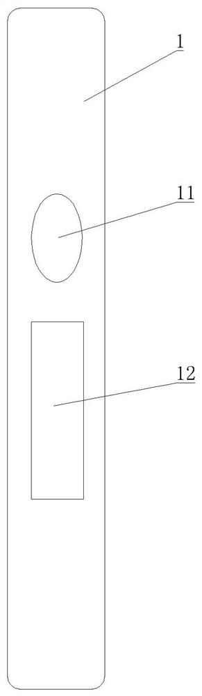
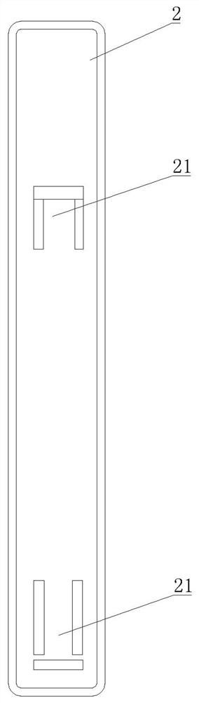
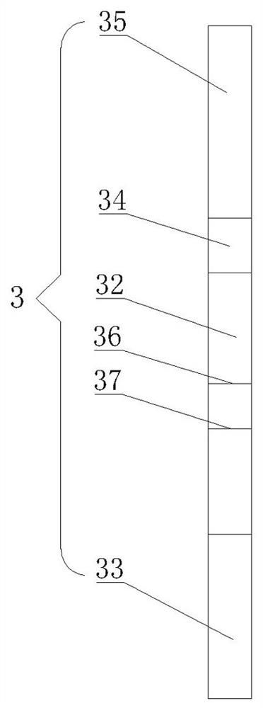
[0035] Embodiment one: as attached figure 1 - attached Figure 4As shown, a Chlamydia pneumoniae antigen detection plate comprises a sealed upper cover 1 and a lower cover 2, the upper cover 1 is provided with a sampling hole 11 and an observation window 12, and the lower cover 2 is provided with a card Slot 21, a test paper strip 3 is placed in the card slot 21, and the test paper strip 3 includes a PVC lath 31, and the PVC lath 31 is provided with a nitrocellulose membrane 32, and the nitrocellulose membrane 32 The position corresponds to the position of the observation window 12, the end of the nitrocellulose membrane 32 away from the sample injection hole 11 is provided with an absorbent paper 33, and the other end of the nitrocellulose membrane 32 is provided with a bonding pad 34 , and the binding pad 34 is completely covered by the upper cover 1 , the free end of the binding pad 34 is provided with a sample pad 35 , and the position of the sample loading hole 11 corres...

Embodiment 2

[0046] Embodiment 2: A Chlamydia pneumoniae antigen detection kit, including the detection plate described in Embodiment 1, and also includes an extraction tube, a disposable sampling swab, and a sample detection solution.

[0047] The sample detection solution is composed of 20mmol / L, Tris-HCL buffer solution with pH of 8, S9 with a mass fraction of 2.5%, Nacl with a mass fraction of 0.9%, Tween with a volume fraction of 0.01%, and Tween with a mass fraction of 0.1%. casein.

[0048] The detection of this kit adopts the immunofluorescence double-antibody sandwich method combined with the amplification system. Its basic principle is the combination of immunochromatography detection technology and amplification system labeling technology. Biotin-labeled Chlamydia pneumoniae antibody and fluorescently-labeled Chlamydia pneumoniae antibody, nitrocellulose membrane 32 coated with avidin and goat anti-mouse quality control line, when the sample contains Chlamydia pneumoniae antigen...


PUM


Abstract

The invention provides a chlamydia pneumoniae antigen detection plate and a detection kit, the chlamydia pneumoniae antigen detection plate comprises an upper cover and a lower cover which are sealed and buckled, the upper cover is provided with a sample adding hole and an observation window, the lower cover is provided with a clamping groove, and a test strip is placed in the clamping groove, the chlamydia pneumoniae antigen detection plate is characterized in that the test strip comprises a PVC (polyvinyl chloride) batten, the PVC batten is provided with a nitrocellulose membrane, and the nitrocellulose membrane is connected with the upper cover. The position of the nitrocellulose membrane corresponds to the position of the observation window, absorbent paper is arranged at one end, far away from the sample adding hole, of the nitrocellulose membrane, a combination pad is arranged at the other end of the nitrocellulose membrane, the combination pad is completely covered by the upper cover, a sample adding pad is arranged at the free end of the combination pad, and the sample adding pad is arranged at the free end of the upper cover. The position of the sample adding hole corresponds to that of the sample adding pad; according to the invention, antigen detection is adopted, the antigen detection can be detected in an incubation period, an acute period or an initial disease course, and the antibody detection is later.

Description

technical field [0001] The invention relates to a detection device for detecting chlamydia pneumoniae antigen. [0002] Specifically, it relates to a chlamydia pneumoniae antigen detection plate and a detection box. Background technique [0003] Chlamydia pneumonia is pneumonia caused by Chlamydia. Chlamydia is divided into Chlamydia trachomatis (CT), Chlamydia pneumoniae (CP), Chlamydia psittaci and Chlamydia familiae. Chlamydia pneumoniae is the most common cause of pneumonia. Chlamydia pneumoniae pneumonia is more common in school-age children, most of which are mild, and the onset is often hidden. There is no gender difference in the infection rate, and it can occur in all seasons. [0004] Chlamydiapneumoniae (Cpn) is a Gram-negative microorganism that parasitizes in cells and is one of the common community-infected respiratory pathogens. Can cause diseases such as pneumonia, pharyngitis, asthma and acute bronchitis. The elderly, infirm, malnourished, and those with...

Claims


Application Information

Patent Timeline
IPC IPC(8): G01N33/569G01N33/558G01N33/533G01N21/64
CPCG01N33/56927G01N33/558G01N33/533G01N21/6428G01N2021/6439G01N2333/295G01N2469/10
Inventor 杨帆李桂芹朱楠楠杨金红李苗苗
Owner 山东康华生物医疗科技股份有限公司
Why Eureka
Try Eureka