
Method for correcting brain tissue deformation in navigation system of neurosurgery

A surgical navigation and neurosurgery technology, applied in the field of medical image processing and application, can solve the problems of complicated intraoperative operation, difficult measurement of cerebrospinal fluid, and inconvenient clinical application, and achieve the effects of convenient clinical application, flexible operation and simple implementation.

Inactive Publication Date: 2005-02-23
FUDAN UNIV
View PDF0 Cites 22 Cited by
  • Summary
  • Abstract
  • Description
  • Claims
  • Application Information

AI Technical Summary

Problems solved by technology

The Image Processing and Analysis research group of Yale University proposed a linear elastic physical model on the IEEE Workshop on Mathematical Methods in Biomedical Image Analysis in November 2001. They used surface displacement as the boundary condition and used dual stereo cameras to obtain the boundary condition. The method of obtaining boundary conditions needs to change the existing navigation equipment to fix the two cameras. At the same time, the correction and synchronization of the two cameras make the intraoperative operation more complicated, so it cannot be conveniently applied in the clinic
The Biomedical Modeling Laboratory of Vanderbilt University published "Model-Updated Image Guidance: Initiai Clinical Experience with Gravity-Induced BrainDeformation" in "IEEE Transactions on medical imaging" (Vol.18, No.10:866-874, 1999.) A theoretical model of consolidation is proposed, but because the model requires physical strength as a boundary condition, that is, the location of the cerebrospinal fluid needs to be obtained during the operation. However, the cerebrospinal fluid is difficult to measure clinically, making this model currently unavailable for clinical application

Method used


Image

  • Method for correcting brain tissue deformation in navigation system of neurosurgery
  • Method for correcting brain tissue deformation in navigation system of neurosurgery
  • Method for correcting brain tissue deformation in navigation system of neurosurgery

Examples


Embodiment 1

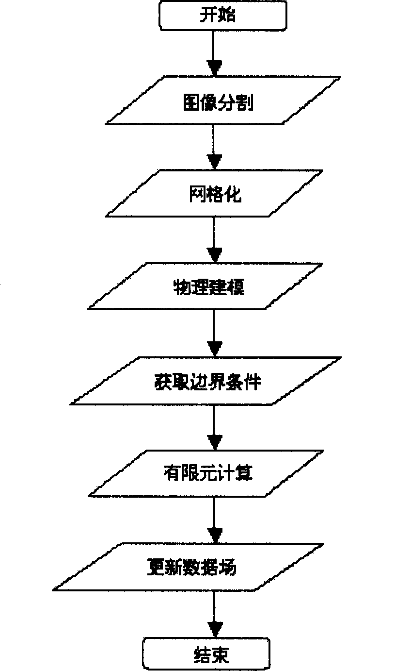
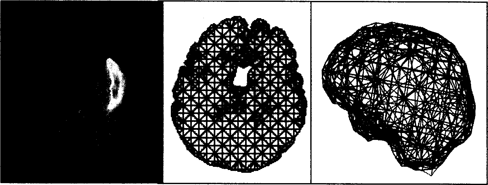
[0054] 1. For the 256×256×48 3D MRI data field, use the 3D automatic segmentation algorithm to obtain brain tissue. The threshold values ​​correspond to the first and second peaks of the fitted Gaussian curve respectively, the corrosion element adopts a spherical element with a radius of 5 pixels, and the expansion element adopts a spherical element with a radius of 6 pixels.

[0055] 2. Using a multi-resolution grid algorithm, the segmented brain tissue is discretized into 18485 tetrahedrons with 5410 nodes. The largest tetrahedron at the boundary is 7.5×7.5×7.5mm 3 (Measured by the size of the hexahedron circumscribed by the tetrahedron), the largest tetrahedron inside is 15×15×15mm 3 ;

[0056] 3. Set the biomechanical property parameters of brain tissue for each unit. Young's modulus=3Kpa, Poisson's ratio=0.45;

[0057] 4. Use coordinate transformation to realize rigid body registration, and obtain the initial position of the cerebral cortex in the LRS space. The trac...


PUM


Abstract

A method for correcting the deformation of cerebral tissue in the navigation system of neurosurgical operation includes such steps as obtaining target tissue (cerebral tissue) by 3D automatic division algorithm based on MRI, generating the lattice of cerebral tissue, assigning the relative biomechanical attribute to each lattice unit, creating physical mode, tracking the movement of exposed cerebral cortex layer, finit element calculating to obtain the deformation for cerebral tissue, and updating the 3D data field before operation by an algorithm to direct the operation.

Description

technical field [0001] The invention belongs to the field of medical image processing and application, and relates to a precision correction method for a surgical navigation system, in particular to a method for correcting brain tissue deformation in a neurosurgery navigation system. Background technique [0002] Brain tissue deformation in clinical surgery is an important factor affecting the accuracy of neurosurgery navigation system. Current solutions include intraoperative image correction such as intraoperative MRI, intraoperative US, etc., and physical model correction. Among them, intraoperative image correction is the method with the highest accuracy, but the disadvantage is that it is expensive and easy to cause intraoperative infection. Therefore, current research mainly focuses on the correction method based on physical models. The method based on the physical model can constrain the movement of brain tissue through the biomechanical properties of the brain tissu...

Claims


Application Information

Patent Timeline
IPC IPC(8): A61B34/10A61B34/20G06F17/00G06T1/00G06T15/00
Inventor 宋志坚刘翌勋李文生王满宁谢震中杜文健
Owner FUDAN UNIV
Learn More