Magnetic separation enzymatic chemical luminous immune detection method of human thyroglobulin antibody

A technology of thyroglobulin and enzymatic chemiluminescence, which is applied in the direction of chemiluminescence/bioluminescence, analysis and measurement devices through chemical reactions of materials, and can solve the problems of unstable radioactive conjugates, short shelf life, and health hazards etc. to achieve the effects of reducing non-specific adsorption, improving sensitivity, and high affinity

Inactive Publication Date: 2006-01-11
北京倍爱康生物技术有限公司
View PDF0 Cites 26 Cited by
  • Summary
  • Abstract
  • Description
  • Claims
  • Application Information

AI Technical Summary

Problems solved by technology

These disadvantages include health hazards, waste disposal problems, short shelf life, instability of radioconjugates, and anti-nuclear pressure from the public (C. Dodeigue, L. Thunus, R. Lejeune. Talanta, 2000, 51: 415-439.)

Method used


Image

  • Magnetic separation enzymatic chemical luminous immune detection method of human thyroglobulin antibody
  • Magnetic separation enzymatic chemical luminous immune detection method of human thyroglobulin antibody

Examples


Embodiment 1

[0041] Example 1: Preparation of secondary antibody enzyme-labeled reagent

[0042] Mix mouse anti-human immunoglobulin A, G, E, M, D, κ chain and λ chain monoclonal antibodies, and concentrate the concentration to 5mg / mL; add 20mmol / L activator N-succinimide 3-(2 -20 μL of pyridyldithiol)propionic acid [SPDP] dimethyl sulfoxide solution, let stand at room temperature for 45 minutes; remove the activator through a Sephadex G25 column, and collect the protein peak; add 500 μL of 24 mg / mL dithiothreose to each ml of activated antibody solution Alcohol (DTT) 0.01mol / L PBS pH7.4 solution, after mixing, let it stand at room temperature for 30 minutes; remove free dithiothreitol through a Sephadex G25 column, and collect protein peaks.

[0043]Dissolve alkaline phosphatase in 2mmol / L EDTA 20mmol / L Tris-HCl pH8.0 solution, the concentration is 5mg / mL; add 0.10mg / mL Traunt reagent (sulfhydryl activating reagent) and let it stand at room temperature for 1 hour; remove free activator t...

Embodiment 2

[0046] Embodiment 2: the preparation of solid phase reagent

[0047] Activate ferric oxide microspheres with a diameter range of 0.1 μm with glutaraldehyde, mix at room temperature for 4 hours, wash with 0.01mol / L PBS pH7.4 buffer three times, and use this solution to suspend at a concentration of 50- 100mg / mL; then, add human thyroglobulin antigen 100μg to each milliliter suspension, mix and incubate at 37°C for 3-8 hours; use an equal volume of 0.01mol / L PBS 5%BSA pH7.4 Block at ℃ for 40 minutes; finally, wash three times with 0.5% BSA 0.02mol / L Tris-HCl pH8.0 buffer solution, and use this solution to prepare a working solution of 8 mg / mL.

Embodiment 3

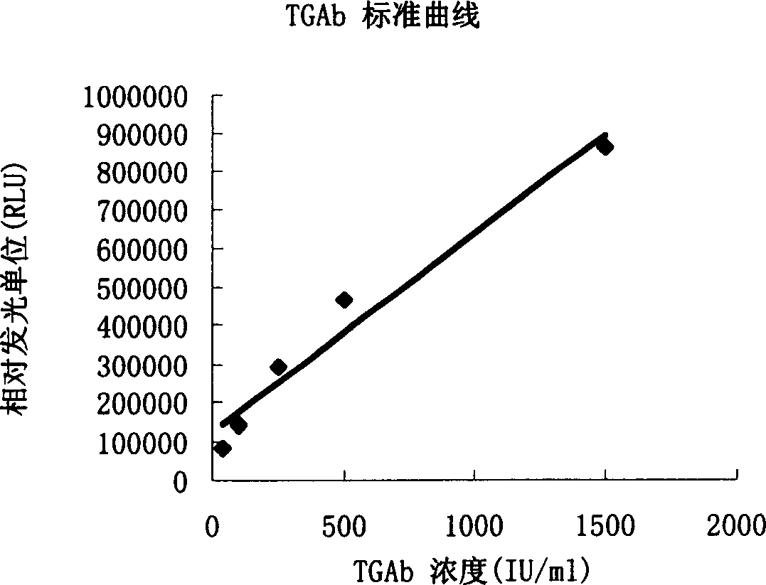
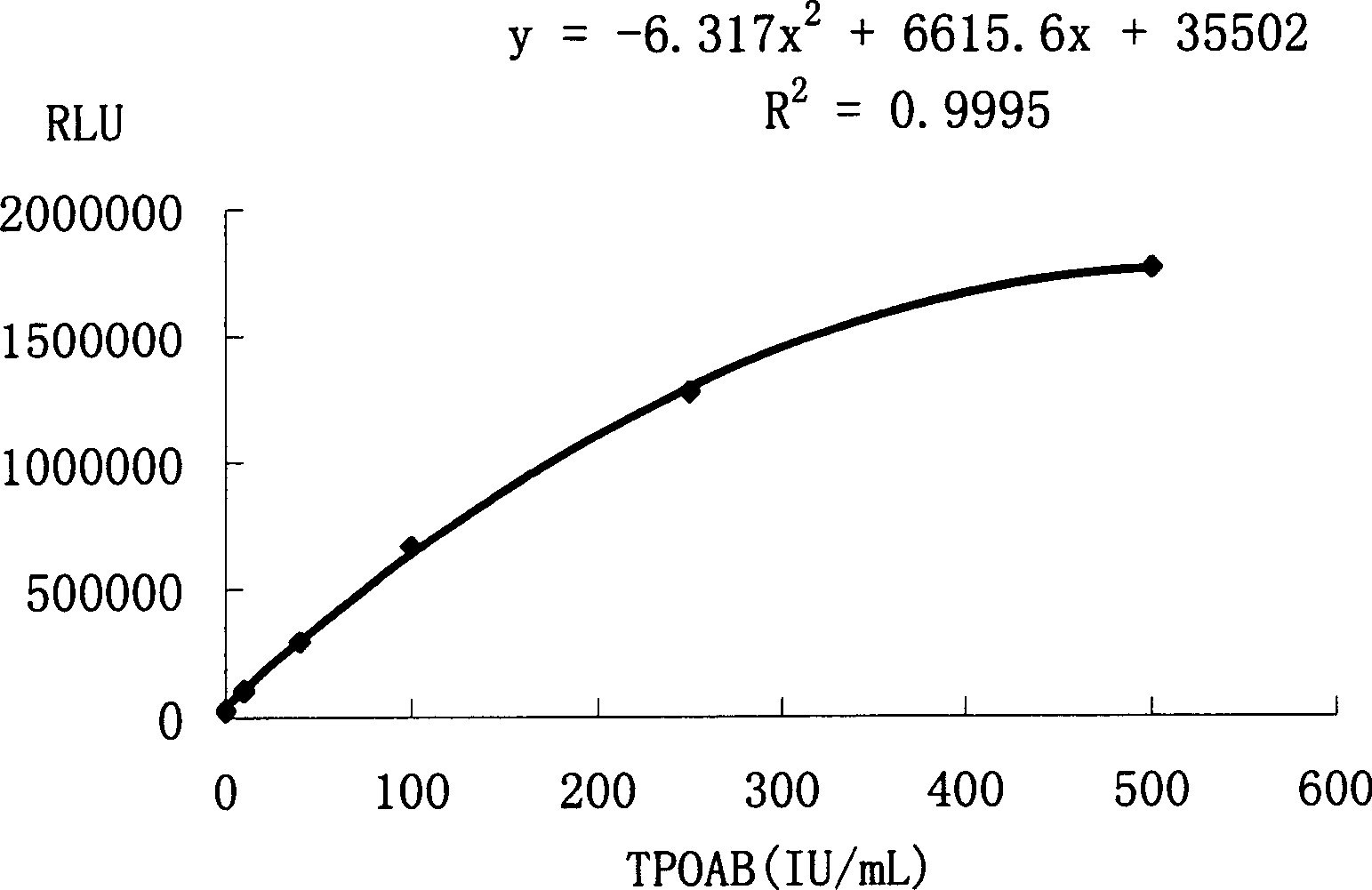
[0048] Example 3: Magnetic separation of human thyroglobulin antibody by enzymatic chemiluminescence immunoassay

[0049] Materials and Instruments

[0050] 1. Standard 0, 40, 100, 250, 500, 1500IU / mL (refer to WHO 1 ST IRP 66 / 387 standard)

[0051] 2. Solid-phase reagent: see Embodiment 2 for details.

[0052] 3. Secondary antibody enzyme-labeled reagent: see Embodiment 1 for details.

[0053] 4. Washing solution 100mL;

[0054] 5. CSPD  Substrate solution: purchased from Applied Biosystems, USA

[0055] 6. The enhancer solution was purchased from Applied Biosystems, USA

[0056] 7. Sample diluent

[0057] 8. 100 serum samples from patients with thyroid autoimmune disease and 100 serum samples from normal people.

[0058] 9. BPCL chemiluminescence detector (manufactured by Institute of Biophysics, Chinese Academy of Sciences).

[0059] 10. Water bath (for 37°C warm bath).

[0060] Reagent preparation:

[0061] The preparation of the standard product is: 2g sodium...


PUM

PropertyMeasurementUnit
diameteraaaaaaaaaa

Abstract

The present invention provides a human thyroglobulin antibody magnetic separation enzymatic chemiluminescent immunoassay method, belonging to the field of immunodetection and analysis technology. Said method includes the following steps: making antigen human thyroglobulin be connected with Fe3O4 mincrosphere surface as solid phase reagent, the diameter of Fe3O4 microsphere is 0.1-5.0 microns, after the self-body antibody human thyroglobulin antibody being in the sample is trapped and two antibodies are labeled by enzyme reagent, the solid phase-antigen-antibody-enzyme-labeled two-antibody sandwiched immune complex can be formed.

Description

technical field [0001] The invention belongs to the technical field of immune detection and analysis, and in particular provides a magnetic separation enzymatic chemiluminescence immunodetection method for human thyroglobulin antibody, which is suitable for magnetic separation enzymatic chemiluminescence detection and analysis of autoantibodies. Background technique [0002] In clinical detection and analysis, due to the low content of many substances in body fluids, it is difficult to meet the detection requirements by using ordinary chemical analysis methods. In 1959, Yalow and Berson used radioactive labeling technology and antigen-antibody immune reaction principle to detect insulin in serum, established a new method of sensitive immunoassay (R.S.Yalow, S.A.Berson.Nature, 1959, 184:1648), and obtained It has been widely used and has become the most popular clinical immunoassay technology developed since the 1960s. But in recent years, the shortcomings of radioimmunity h...

Claims


Application Information

Patent Timeline
Patent Type & Authority Applications(China)
IPC IPC(8): G01N33/538G01N21/76
Inventor 刘振世周昊王法龙王东张波谢晶陈海生胡晓焱张小平孙海涛张玉庆杨祥良
Owner 北京倍爱康生物技术有限公司
Why Eureka
Try Eureka