Living body observing apparatus, intraoral imaging system, and medical treatment appliance

A technology of living body observation and camera device, which is applied in oral mirrors, diagnosis, application, etc., can solve the problems of undisclosed adjustment of excitation light and white light, and achieve the effect of improving treatment accuracy

Inactive Publication Date: 2007-04-25
MORITA MFG CO LTD
View PDF3 Cites 19 Cited by
  • Summary
  • Abstract
  • Description
  • Claims
  • Application Information

AI Technical Summary

Problems solved by technology

[0014] In Patent Document 3, although white light can be irradiated in addition to the excitation light (laser), there is no way to simultaneously irradiate two kinds of light and obtain a clear fluorescent image for identifying a lesion in a clear image of normal tissue. There are some disclosures or hints about the technical features, and there is no disclosure of the technical idea of ​​obtaining an appropriate image corresponding to the detection purpose by appropriately adjusting the relative illuminance of the excitation light and the white light
Moreover, the dental caries detection device in this patent document also uses a light guide device to guide the radiation from the irradiation device, and there is no description that the generator of excitation light and illumination light is integrated with the camera, and it is still enlarged. device

Method used


Image

  • Living body observing apparatus, intraoral imaging system, and medical treatment appliance
  • Living body observing apparatus, intraoral imaging system, and medical treatment appliance
  • Living body observing apparatus, intraoral imaging system, and medical treatment appliance

Examples


Embodiment 1

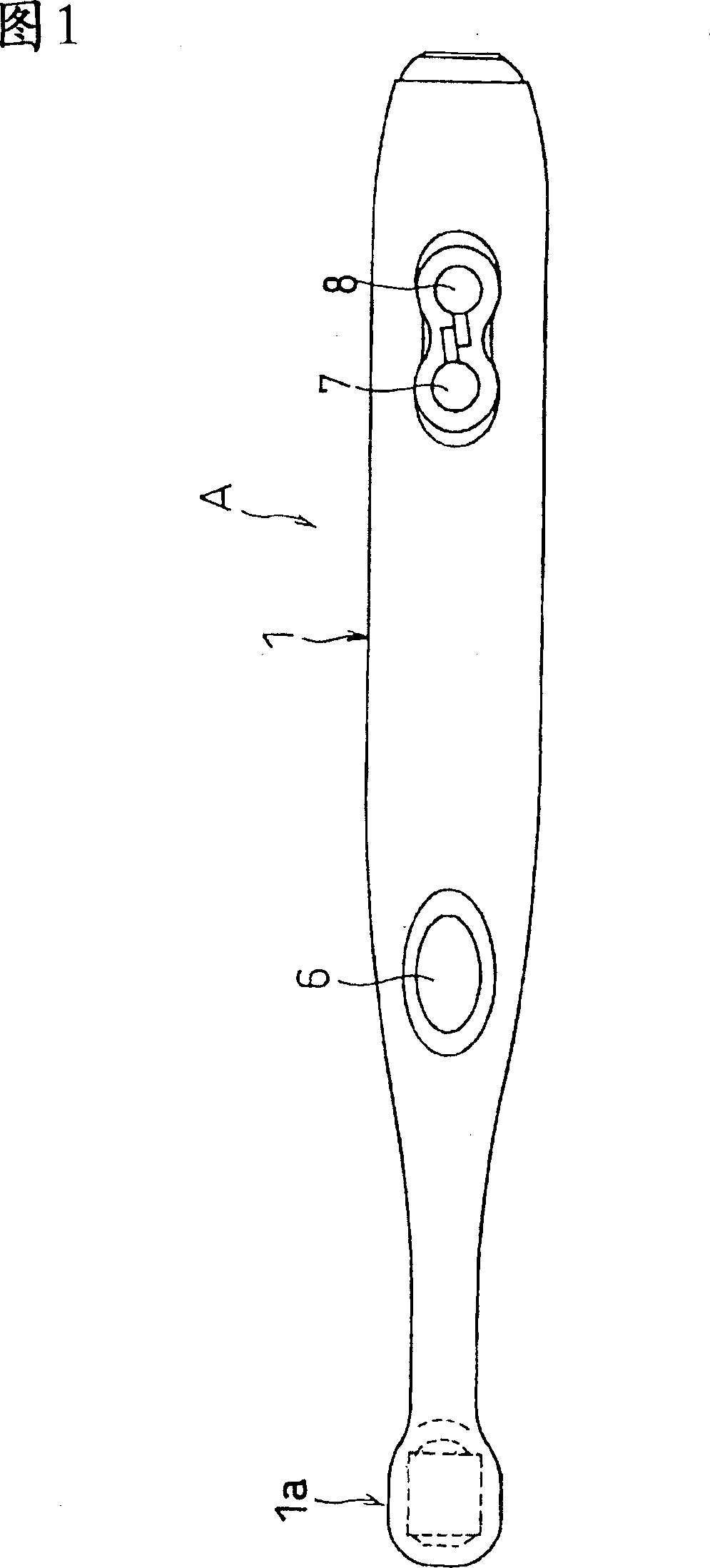
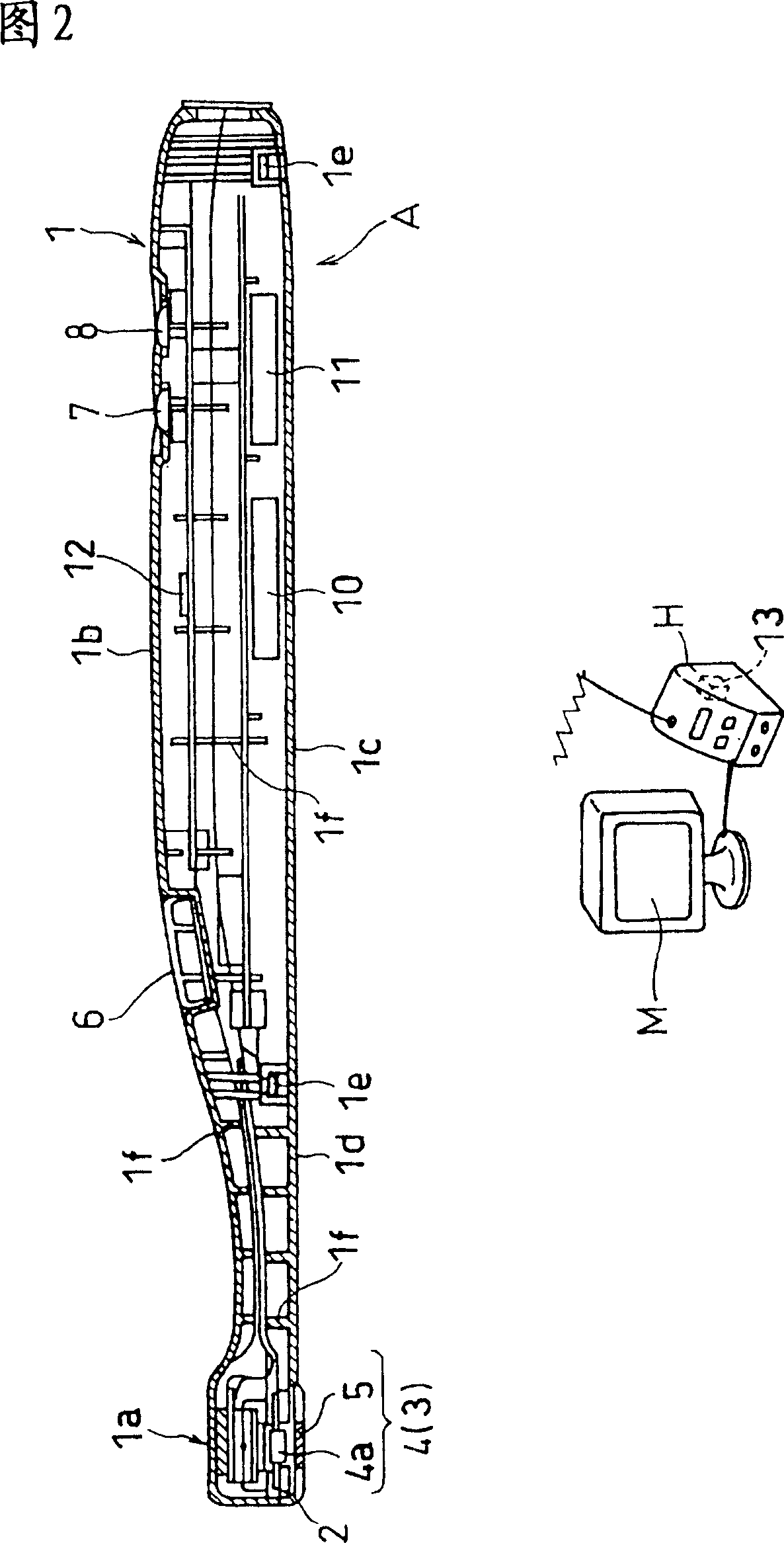
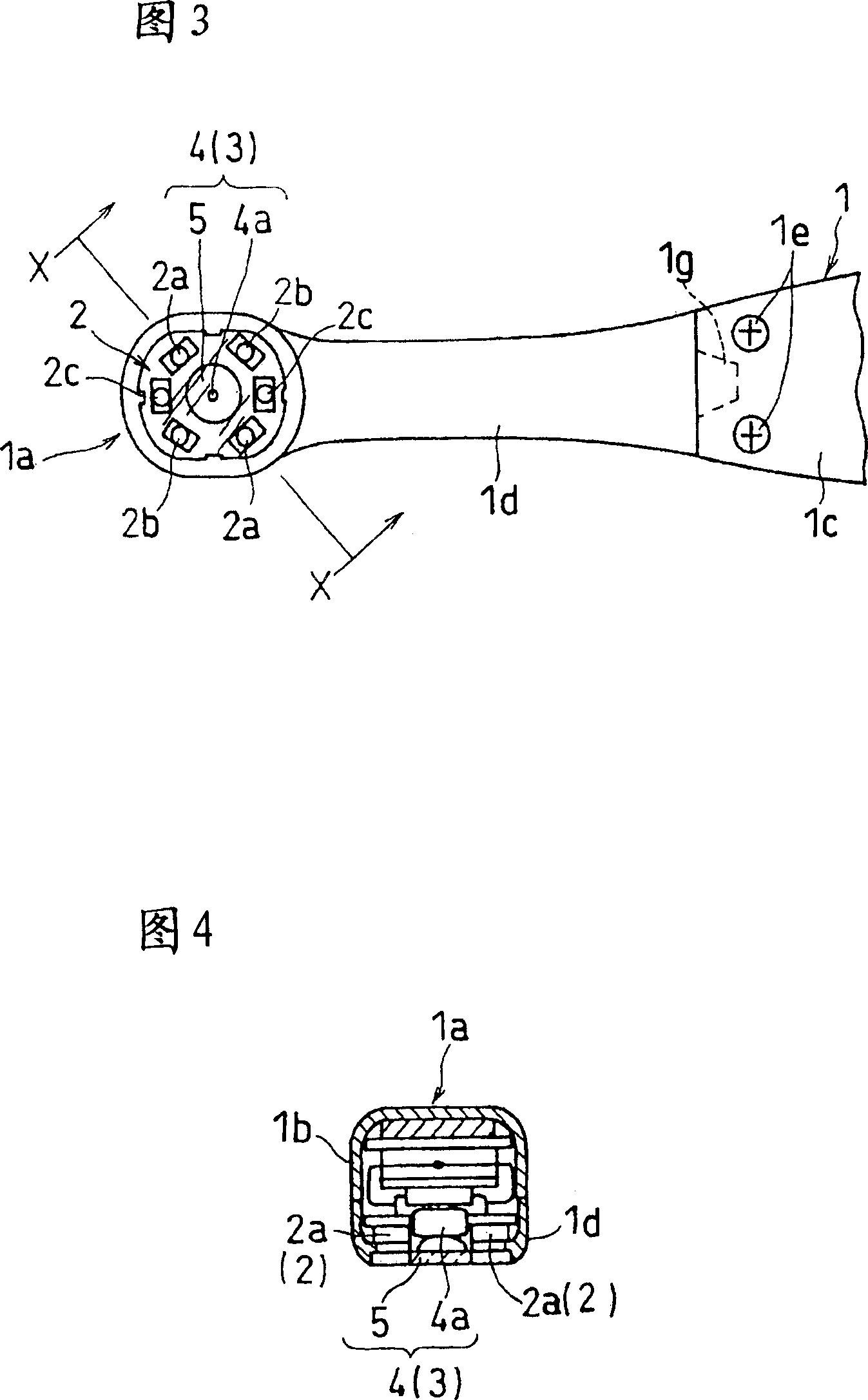
[0082] Fig. 1 is a plan view showing an example in which the living body observation instrument of the present invention is embodied in an intraoral imaging device, Fig. 2 is a longitudinal sectional view along the longitudinal direction of the imaging device, and Fig. 3 is an enlarged bottom view of the top side of the main body Fig. 4 is a longitudinal sectional view taken along line X-X of Fig. 3 . The intraoral imaging device A in the figure has an imaging device 4 serving as an irradiation device 2 and an observation unit 3 on the top side portion 1a of a dental handpiece-shaped body (casing) 1 freely supported by fingers. This intraoral imaging device A is mainly suitable for diagnosing dental caries, defective parts, lesion parts, tartar, tartar, or biofilm adhesion degree of the teeth in the oral cavity. H (refer to Figure 2) transmits wireless signals and prints out the captured images. In addition, it is also possible to provide a zoom structure or an automatic focu...

Embodiment 2

[0102] The intraoral imaging device A1 in FIG. 10 is an example showing that the irradiation device is detachable. The intraoral imaging device A1 in the figure has an irradiation device (LED) 2 for irradiating excitation light and illumination light to the observation target part on the top side part 1a of the main body casing 1 that can be freely supported by fingers, and The imaging device 4 as the observation unit 3 . The imaging device 4 is composed of the same solid-state imaging element 4 a and a light-receiving filter 5 as described above. The light-receiving filter 5 transmits the fluorescence radiated from the lesion of the observation target site and the illumination light reflected from the periphery of the lesion, and guides the light to the solid-state imaging device 4a. Moreover, the irradiation device 2 can be freely attached and disassembled relative to the top side part 1a.

[0103] That is, an annular support portion 5a for supporting the light-receiving f...

Embodiment 3

[0108]The intraoral imaging device A2 in FIG. 11 shows an example in which the imaging device has an optical path conversion device. In the intraoral imaging device A2 in the figure, the solid-state imaging element 4a constituting the imaging device 4 serving as the observation section 3 is installed in the top side portion 1a of the main body housing 1 with its optical axis along the longitudinal direction of the main body housing 1. , and, as an optical path conversion device, a mirror (or prism) 18 is installed on the top side inner surface of the top side part 1a at an angle of about 45 degrees with respect to the above-mentioned optical axis, and the inner tube between the mirror 18 and the solid-state imaging element 4a The part becomes the light guide path 19 for the imaging light. A relay lens 20 and a relay lens 21 movable along the optical axis are provided on the light guide path 19 . An optical system for forming an optical image on the solid-state imaging device ...


PUM


Abstract

A medical living body observation apparatus, an intraoral photography system and a medical treatment appliance which can be used conveniently by a general user at home as well as a technician, e.g. a dentist or a physician. There is provided an irradiation means (2) comprising an exciting light emission section (2a) emitting an exciting light for causing a lesion to radiate fluorescence, and an illumination light emission section (2b) emitting a light for illuminating the periphery of the lesion. The irradiating means (2) can simultaneously irradiate the illumination light and the exciting light.

Description

technical field [0001] The present invention relates to the diagnosis of dental caries, defects, lesions, or adhesion of calculus or tartar, root canals, gums, cheeks, and tongue lesions in the oral cavity, or diagnosis in the field of otolaryngology. It further relates to an intraoral imaging device capable of clearly viewing a lesion and taking an image. In addition, it relates to a living body observation instrument, an intraoral imaging device, and a medical diagnostic tool that can observe the adhesion of calculus or the progress of dental caries in the oral cavity of oneself or family members, and even observe the skin, etc. at home. Background technique [0002] For example, an intraoral imaging device used for diagnosing the oral cavity needs to be placed in the mouth for manipulation, so the imaging device needs to be reduced in size. That is, such an intraoral imaging device has traditionally been a structure in which a light source or a CCD imaging device for irr...

Claims


Application Information

Patent Timeline
Patent Type & Authority Applications(China)
IPC IPC(8): A61B1/00A61B1/24A61B5/00
CPCA61B1/0684A61B5/0088A61B1/24A61B1/0615A61B1/043A61B1/0692A61B1/0623A61B1/06A61B1/0676A61B1/0605A61B1/00
Inventor 大川真一木野健二的场一成
Owner MORITA MFG CO LTD
Why Eureka
Try Eureka