Evolved immunoglobulin binding molecule, and its preparation method and uses

An immunoglobulin and binding molecule technology, applied in hybrid immunoglobulins, biochemical equipment and methods, botanical equipment and methods, etc., can solve the problem that the functional characteristics of LD5 are not further elaborated, and achieve improved sensitivity and accuracy. High performance, good effect, low cost effect

Inactive Publication Date: 2007-05-23
SECOND MILITARY MEDICAL UNIV OF THE PEOPLES LIBERATION ARMY
View PDF0 Cites 8 Cited by
  • Summary
  • Abstract
  • Description
  • Claims
  • Application Information

AI Technical Summary

Problems solved by technology

Specifically: cloning the single Ig-binding domains of SpA and PpL, splicing the single-domain DNA fragments randomly in vitro, and then applying phage display technology to express these recombinant molecules on the surface of phage, and constructing a random combination of single-domains of SpA and PpL Phage display library, using human Ig as bait to perform affinity screening on the library, the result is a new type of Ig composed of the B3 domain of PpL and the D, A, B, C (mainly D) domains of SpA arranged in intervals Binding molecular form (MDPL-MDPA)n(MDPL, mono-domain of protein L; MDPA, mono-domain of protein A), which does not exist in natural IBP and has not been reported in recombinant IBP molecules, therefore It is a new molecular structure form of IBP. Since it is obtained by means of molecular evolution, we collectively refer to these molecules as evolved Ig-binding molecules, and evolved Ig-binding molecules LD5 (L-D-L-D-L; L is the B3 domain of PpL, and D is the D domain of SpA) is the most representative dominant clone among them. Progress in Biochemistry, Volume 32, Issue 6, 2005, pages 535-543 have a detailed description, but this article does not further elaborate on the various functional properties of LD5

Method used


Image

  • Evolved immunoglobulin binding molecule, and its preparation method and uses
  • Evolved immunoglobulin binding molecule, and its preparation method and uses
  • Evolved immunoglobulin binding molecule, and its preparation method and uses

Examples


Embodiment 1

[0138] Example 1 A method for prokaryotic preparation of novel evolved immunoglobulin binding molecules

[0139] 1. cDNA sequence and cloning of novel evolved immunoglobulin binding molecules

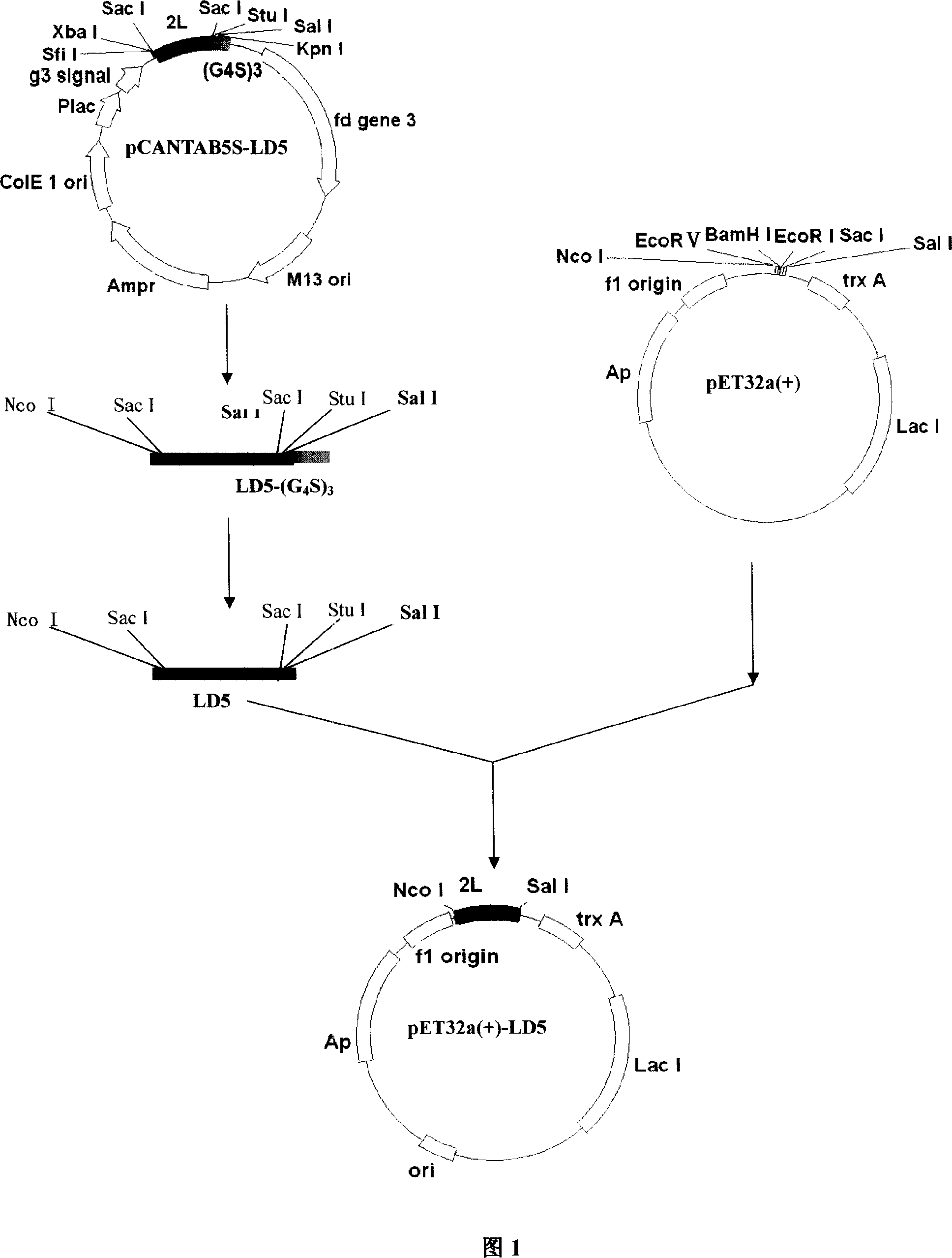
[0140] The recombinant phagemid vector pCANTAB5S-LD5, which contains the cDNA sequence clone encoding the evolved immunoglobulin binding molecule LD5 (L-D-L-D-L; L is the B3 domain of PpL, and D is the D domain of SpA), was obtained through molecular evolution screening and prepared The specific steps of pCANTAB5S-LD5 have been fully described in "Phage Display of SpA and PpL Ig Binding Single Domain Random Combinatorial Library and Affinity Screening", Progress in Biophysics and Biochemistry, Volume 32, Issue 6, Pages 535-543, 2005 Open, that is, the 27# / 44# of the third round of screening and the 4# / 33# of the fourth round of screening in Table 4 in the literature. The cloning vector, its cDNA sequence is shown in SEQ ID No.1, and its amino acid sequence is shown in SEQ ID No...

Embodiment 2

[0173] Example 2 Binding of novel evolved immunoglobulin binding molecules to various antibodies and antibody fragments

[0174] 1. Binding to human polyclonal IgG:

[0175] 1.1 Human polyclonal IgG was purchased from Sigma, and PpL and SpA were purchased from Sigma.

[0176] 1.21 mg / ml human polyclonal IgG was dialyzed against PBS (pH 7.2). Take 50 μL of 3 mg / ml long-arm activated biotin (purchased from PIERCE Company) and add 1 mL of IgG (1 mg / mL), shake slightly at room temperature for 4 hours, then add a dialysis bag and dialyze overnight in PBS at 4 °C, add an equal amount of glycerol after the dialysis Store at -20°C.

[0177] 1.3 Adjust the concentration of LD5, PpL and SpA to 1 mg / ml, then dilute with carbonate buffer (pH9.6) 1:200 and coat a 96-well plate. Each protein is coated with 3 rows of duplicate wells. After 24 hours at 4°C, Wash 4-5 times with PBST, block with blocking solution (2% BSA 0.05% TWEEN-20) and wash the plate at 37°C for 1 hour. Biotin-lab...

Embodiment 3

[0201] Example 3 Demonstration of the Two-Site Binding Mode of the Novel Evolved Immunoglobulin Binding Molecule Kappa Light Chain and VH3 Heavy Chain

[0202] SpA has a strong affinity with the Fc segment of IgG, and at the same time SpA can also bind to the VH region belonging to the VH3 gene family (SassoEH, Silverman GJ, Mannik M. Human IgA and IgG F(ab')2 that bind to staphylococcalSpA belong to the VHIII subgroup. J Immunol. 1991; 147: 1877-1883). PpL binds immunoglobulins by interacting with Ig light chains, primarily subtypes 1, 3, and 4 of the kappa light chain (Nilson BH, Solomon A, BjorckL, et al. PpL from peptostreptococcus magnus binds to the kappa light chain variable domain; J Biol Chem. 1992;267(4):2234-9). According to the binding experiments between LD5 and various Ig molecules, LD5 has a higher affinity to IgG Fab IgM IgA than SpA and PpL. It is speculated that LD5 has dual-site binding properties to the VH3 heavy chain and κ light chain variable regions. ...


PUM

PropertyMeasurementUnit
molecular weightaaaaaaaaaa
Sensitivityaaaaaaaaaa

Abstract

The invention relataes to novel evolutionary immunoglobulin binding molecules, as well as their preparation methods and applications. The invention discloses separated evolutionary immunoglobulin binding molecules, which are proteins with amino acid sequences as shown by SEQ ID NO : 1, or conservative variant proteins with immunoglobulin binding activity. The invention also discloses the gene encoding, genetic engineering preparation methods and applications of immunoglobulin binding molecules. The disclosed immunoglobulin molecule broad-spectrum combined with various immuneglobulin shows high immunoglobulin whole molecule binding activity, and can be used in large-scale purification of genetic engineering antibodies, purification of natural antibodies and monoclonal antibodies, enzyme-linked immunosorbent assay, and immuno-chromatography and immunohistochemical methods for immune antibody detection and diagnosis.

Description

technical field [0001] The present invention relates to a recombinant immunoglobulin binding molecule, in particular to an immunoglobulin binding molecule obtained by rearranging the Ig binding domain of Staphylococcus aureus protein and the surface protein of Streptococcus majoris, its preparation method and application. Background technique [0002] Immunoglobulin-binding molecules (Ig binding proteins, IBP) are a class of proteins expressed on the surface of bacteria that can bind to specific parts of host Ig, regulate the host's immune response to bacteria, and are one of the important pathogenic factors of bacteria. Three main types of IBP have been studied the most: Staphylococcus aureus protein A (SpA), human streptococci (groups C and G) protein G (SpG) and surface protein L (PpL) of some Peptostreptococci magna. [0003] SpA has a molecular weight of about 57KDa and is composed of about 524 amino acids. Its extracellular part contains five Ig-binding domains with a ...

Claims


Application Information

Patent Timeline
Patent Type & Authority Applications(China)
IPC IPC(8): C07K16/46C12N15/31C12N15/70C12N1/21G01N33/53G01N33/577
Inventor 潘卫蒋少华徐容贾建安沈毅珺杨华王锦红陈秋莉何俊陈璐
Owner SECOND MILITARY MEDICAL UNIV OF THE PEOPLES LIBERATION ARMY
Why Eureka
Try Eureka