
Tim protein-bound carrier, methods for obtaining, removing and detecting extracellular membrane vesicles and viruses using said carrier, and kit including said carrier

a technology carrier proteins, which is applied in the field of tim protein-bound carriers, can solve the problems of difficult separation of extracellular membrane vesicles from hdl having the same density, difficult to obtain highly pure extracellular membrane vesicles, etc., and achieves high purity and high sensitivity

Active Publication Date: 2020-10-06
OSAKA UNIV +1
View PDF9 Cites 2 Cited by
  • Summary
  • Abstract
  • Description
  • Claims
  • Application Information

AI Technical Summary

Benefits of technology

The method allows for the efficient and sensitive detection and removal of extracellular membrane vesicles and viruses in high purity, maintaining their intact state, overcoming the limitations of existing techniques.

Problems solved by technology

However, with this method, it is difficult to obtain the highly pure extracellular membrane vesicles, because protein complexes or aggregates, lipoproteins, such as HDL, and the like, contained in a sample are also co-precipitated, other than the extracellular membrane vesicles.
Although it is possible to separate the protein complexes or the aggregates by density fractionation of the precipitate fraction obtained by the ultracentrifugal separation treatment by a sucrose density gradient method, it is difficult to separate them from the HDL having the same density.
In addition, it is difficult to process multiple samples at the same time, since this method requires ultracentrifugal separation treatment.
In addition, expensive equipment is required for ultracentrifugal separation treatment.
(NON-PATENT LITERATURE 1), however, there is a problem that purity of the resulting extracellular membrane vesicles is further lower as compared with a method by the ultra-centrifugal separation treatment.
These methods have such problems that only the extracellular membrane vesicles having the surface antigenic protein for the antibody can be obtained; yield of the extracellular membrane vesicles is small; it is difficult to obtain the extracellular membrane vesicles in an intact state (that is, while maintaining an original state of function of the extracellular membrane vesicles) because it is necessary to use a surfactant, an acidic buffer, or the like, in order to elute the extracellular membrane vesicles from the antibody; and the like, although the high purity extracellular membrane vesicles can be obtained.
However, in this method, it is difficult to obtain the high purity viruses, because the protein complexes and the aggregates, the lipoproteins, such as HDL, and the like, contained in the sample are co-precipitated other than the viruses.
Although it is possible to separate the protein complexes or the aggregates by density fractionation of the precipitate fraction obtained by the ultracentrifugal separation treatment, by a sucrose density gradient method, it is difficult to separate them from the aggregates having the same density.
In addition, there is also a problem that activity of the viruses is lowered by the ultracentrifugal separation treatment (NON-PATENT LITERATURE 2).
In addition, this method is difficult to process multiple specimens at the same time, because it requires the ultracentrifugal separation treatment.
In addition, an expensive machine is required for the ultracentrifugal separation treatment.
In addition, condition setting may be difficult, in some cases, and thus it is difficult to apply it to purification of all viruses.
However, this method has difficulty in complete removal of the extracellular membrane vesicles, thus resulting in remaining of the extracellular membrane vesicles not completely removed, in the sample.
However, in also this method, there was difficulty to completely remove the extracellular membrane vesicles, due to remaining of the extracellular membrane vesicles not completely removed, in the sample.
In addition, there was the case of inducing a problem on bioactivity, or the like, of the extracellular membrane vesicles, by the reagent, because of contamination of the reagent added into the sample.
There has been known the same method also as a method for removing the viruses and there was the same problem as in the method for removing the extracellular membrane vesicles.
However, the minimum detection sensitivity of the sandwich ELISA system using the antibody that detects an ordinary color development signal has been reported to be about 3 μg in purified exosomes, and sufficient sensitivity for measuring a body fluid sample, such as serum, has not been obtained (NON-PATENT LITERATURE 4).
The same method has been known, also as the method for detecting the viruses, and sufficient sensitivity for detection of the viruses has also not been obtained.
However, in the usual method for capturing the extracellular membrane vesicles using a carrier to which the antibody against the surface antigen of the extracellular membrane vesicles was immobilized, sufficient sensitivity has not been obtained; and thus direct detection from the sample, such as culture supernatant or body fluid, has been difficult, and the detection from the sample after concentrating or purifying the extracellular membrane vesicles has been required.

Method used


Image

  • Tim protein-bound carrier, methods for obtaining, removing and detecting extracellular membrane vesicles and viruses using said carrier, and kit including said carrier
  • Tim protein-bound carrier, methods for obtaining, removing and detecting extracellular membrane vesicles and viruses using said carrier, and kit including said carrier
  • Tim protein-bound carrier, methods for obtaining, removing and detecting extracellular membrane vesicles and viruses using said carrier, and kit including said carrier

Examples


experimental example 1

Fusion Type Tim-4 Protein

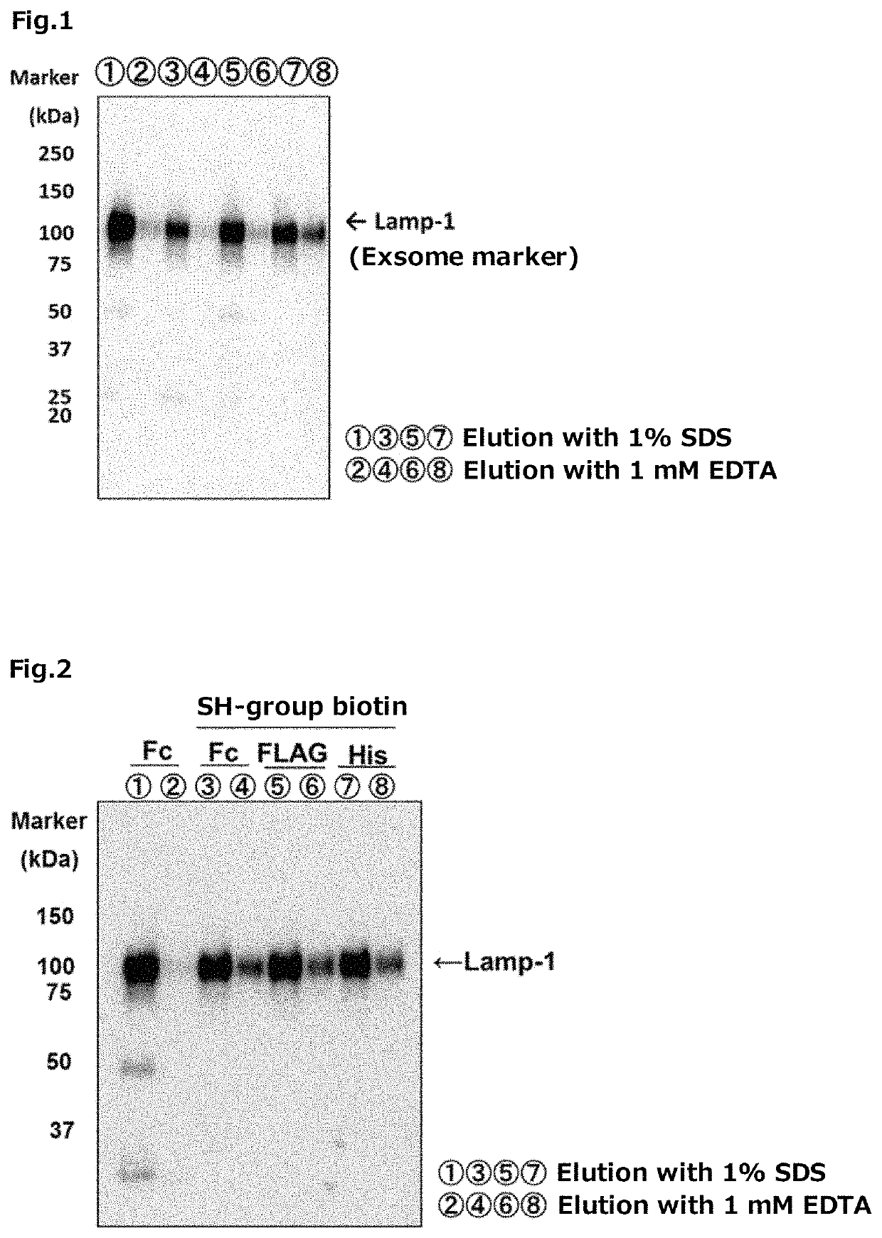
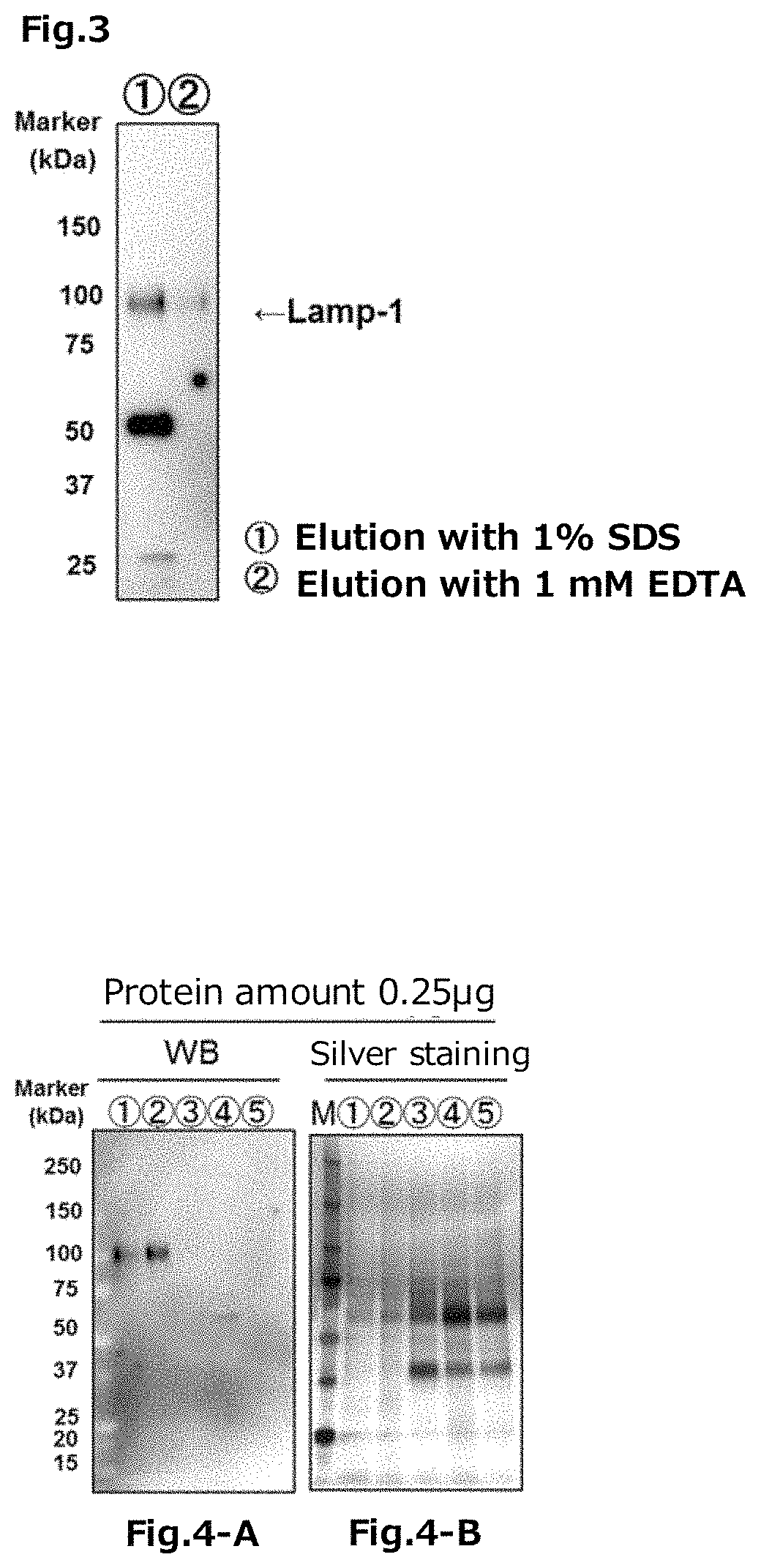
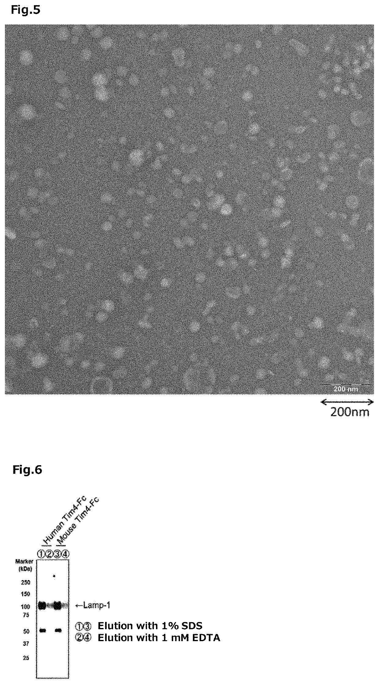
[0351]The Fc tag fusion type Tim-4 protein was prepared by the following method.

[0352]First, a vector was constructed for expression of the Fc tag fusion type Tim-4 protein in which a cDNA (SEQ ID NO: 26) encoding the N-terminal 1 to 273 amino acid region of a mouse-derived Tim-4 protein was incorporated into the Sall-EcoRV site of a pEF-Fc vector (hereinafter it may be abbreviated as “the pEF-Tim-4-Fc”, in some cases).

[0353]On the other hand, 293 T cells (RIKEN BRC) were each cultured for 1 day using 25 sheets of 150 mm dishes for cell culture, using 20 mL of DMEM (produced by Nacalai Tesque, Inc.) containing 10% FBS (produced by BioWest Co., Ltd.). Thereafter, for each dish, 25 mL of DMEM containing 10% FBS was replaced with 25 mL of FBS-free DMEM.

[0354]Thereafter, 20 μg of the pEF-Tim-4-Fc was transfected into each of 293 T cells, using Polyethylenimine “MAX” (produced by Polysciences Inc.), according to a usual method. After transfection, the 293 T cells...

experimental example 2

g Fusion Type Tim-4 Protein

[0358]The FLAG tag fusion type Tim-4 protein was prepared by the following method.

[0359]First, a FLAG tag fusion type mouse-derived Tim-4 protein cDNA (a cDNA encoding an amino acid sequence fused with 1×FLAG at the C-terminal of the N-terminal 1 to 273 amino acid region of the mouse-derived Tim-4 protein, SEQ ID NO: 33 (containing a stop codon (taa) and a nucleotide sequence encoding 1×FLAG, in this regard, however, excluding the restriction enzyme site), produced by Fasmac Co., Ltd.), was incorporated in an XhoI / BamHI site of a pCAG-Neo vector (produced by Wako Pure Chemical Industries, Ltd.) to construct a vector for expressing the FLAG tag fusion type Tim-4 protein (hereinafter it may be abbreviated as “the pCAG-Tim-4-FLAG”, in some cases).

[0360]On the other hand, the 293 T cells (RIKEN BRC) were cultured for 1 day using 50 mL DMEM (produced by Wako Pure Chemical Industries, Ltd.) containing 10% FBS (produced by Biosera) in a 225 cm2 flask by seeding, ...

experimental example 3

Fusion Type Tim-4 Protein

[0363]The His tag fusion type Tim-4 protein was prepared by the following method.

[0364]The His tag fusion type mouse-derived Tim-4 protein cDNA (a cDNA encoding an amino acid sequence fused with 6×His tag at the C-terminal of the N-terminal 1 to 273 amino acid region of the mouse-derived Tim-4 protein, SEQ ID NO: 34 (containing the stop codon (tga) and a cDNA encoding 6×His tag) was incorporated in the XhoI / BamHI site of the pCAG-Neo vector (produced by Wako Pure Chemical Industries, Ltd.) to construct a vector for expressing the His tag fusion type Tim-4 protein (hereinafter it may be abbreviated as “the pCAG-Tim-4-His”, in some cases).

[0365]On the other hand, the 293 T cells (RIKEN BRC) were cultured for 1 day using 50 mL of DMEM (manufactured by Wako Pure Chemical Industries, Ltd.) containing 10% FBS (produced by Biosera) in a 225 cm2 flask. Thereafter, 60 μg of the pCAG-Tim-4-His was transfected into the 293 T cells using Lipofectamine 2000 (produced by ...


PUM

PropertyMeasurementUnit
particle sizeaaaaaaaaaa
pHaaaaaaaaaa

Abstract

The invention provides a carrier and a method for obtaining, removing, or detecting extracellular membrane vesicle or virus present in a sample. In particular, the invention provides (a) a carrier (a Tim carrier) on which a protein (a Tim protein), selected from a T-cell immunoglobulin and mucin domain-containing molecule-4 (a Tim-4) protein, a Tim-3 protein, and a Tim-1 protein, is bound; (b) a method for obtaining the extracellular membrane vesicle or the virus in the sample; (c) a method for removing the extracellular membrane vesicle or the virus in the sample; (d) a method for detecting the extracellular membrane vesicle or the virus in the sample; (e) a kit for capturing the extracellular membrane vesicle or the virus, comprising the Tim carrier; and f) a kit for capturing the extracellular membrane vesicle or the virus, comprising a reagent containing the Tim protein and a reagent containing the carrier.

Description

CROSS-REFERENCE TO RELATED APPLICATIONS[0001]This patent application is the U.S. national phase of International Patent Application No. PCT / JP2015 / 083505, filed Nov. 27, 2015, which claims the benefit of Japanese Patent Application No. 2014-246876, filed on Dec. 5, 2014, which are incorporated by reference in their entireties herein.INCORPORATION-BY-REFERENCE OF MATERIAL ELECTRONICALLY SUBMITTED[0002]Incorporated by reference in its entirety herein is a computer-readable nucleotide / amino acid sequence listing submitted concurrently herewith and identified as follows: 56,202 bytes ASCII (Text) file named “728241ReplacementSequenceListing.txt,” created Sep. 12, 2017.TECHNICAL FIELD[0003]The present invention relates to a Tim protein-binding carrier, a method for obtaining, removing and detecting extracellular membrane vesicles and viruses using the carrier, as well as a kit comprising the carrier.BACKGROUND ART[0004]It has been known that the extracellular membrane vesicles contain pr...

Claims


Application Information

Patent Timeline
Patent Type & Authority Patents(United States)
IPC IPC(8): G01N33/50G01N33/543G01N33/68C12N15/09C12N7/02C07K14/705C07K17/02C07K1/14C07K17/00G01N15/00G01N15/10G01N15/14G01N33/53
CPCC07K17/00G01N33/5076C07K1/14G01N33/5432C12N7/02C12N15/09C07K14/70503G01N33/6842C07K17/02G01N2015/1006C12N2760/16151G01N33/53C12N2760/18551G01N2015/149C07K2319/40G01N2015/008G01N2333/70503G01N15/149G01N2015/016
Inventor NISHIBU, TAKAHIROIMAWAKA, NAOKONARUSE, KENHANAYAMA, RIKINARI
Owner OSAKA UNIV
Learn More