
Methods of treating cellulite and subcutaneous adipose tissue

a cellulite and subcutaneous adipose tissue technology, applied in the field of cellulite and subcutaneous adipose tissue treatment, can solve the problems of non-adipose cells and tissues being damaged, side effects typically subside, application of these technologies is likely to have potential safety issues, etc., to reduce undesired side effects, reduce the volume of subcutaneous adipose tissue, and reduce the appearance of cellulite

Active Publication Date: 2022-01-25
SOLITON
View PDF226 Cites 0 Cited by
  • Summary
  • Abstract
  • Description
  • Claims
  • Application Information

AI Technical Summary

Benefits of technology

This approach allows for shorter treatment sessions with minimal side effects, inducing significant inflammation and fat reduction while maintaining tissue integrity, thus enhancing the effectiveness and practicality of fat reduction procedures.

Problems solved by technology

Excess body fat, localized adiposity, and cellulite represent important social problems.
While both technologies result in adipose tissue destruction, the application of these technologies is likely to have potential safety issues because of the cavitation or thermal affects.
These cavitation or thermal affects may even cause damage to non-adipose cells and tissues.
Other treatment modalities, such as lasers, radiofrequency and focused ultrasound, affect fat cells and may affect other adjacent tissue in a way that is not comparable to the CoolSculpting method of Cryolipolysis®.” While side effects such as transient local redness, bruising and numbness of the skin are common following the cryolipolysis treatment, the company claims the these side effects typically subside over time.
While the use of cryolipolysis to induce an inflammatory response that results in an adipose tissue volume reduction is an improvement over prior art approaches, it is still less than ideal.
One problem with using cryolipolysis to induce inflammation is the time it takes to administer the cryolipolysis treatment (i.e., cooling the adipose tissue).
Another problem, during these long cryolipolysis treatments, the patient is limited on making any movements, which makes the treatment unpleasant.
Additionally, a major problem for the physician or spa owner who is treating the patient, the required long treatments limits the throughput of patients that can be seen which has a real impact on the practice revenues.
The combination of the two procedures causes the programmed death and slow resorption of destroyed adipocytes.”
As a result, the fundamental problems, as discussed above, related to the cryolipolysis treatment has not changed.

Method used


Image

  • Methods of treating cellulite and subcutaneous adipose tissue
  • Methods of treating cellulite and subcutaneous adipose tissue
  • Methods of treating cellulite and subcutaneous adipose tissue

Examples


example 1

Adipose Tissue Inflammation

[0095]A study was undertaken to evaluate the induction of inflammation in subcutaneous fat using high-frequency shockwave. A Gottingen minipig (˜30 Kg) was anesthetized. The mid-ventral sites were prepared by removing the skin hair here using hair clippers and then razor. High-frequency shockwaves were then applied to the two treatment sites. Following the high frequency shockwave treatment, and 48 hours post treatment, biopsies were taken of treatment sites using 3 mm circular punch biopsy instruments. Tissue samples were placed in buffered formalin for microscopic examination.

[0096]The high frequency shockwave treatment protocols are shown in Table 1. The probe had a 30 mm diameter shockwave outlet window and was configured to generate electrohydraulic shockwaves. All five sites that were treated using different high frequency shockwave settings demonstrated inflammation in the subcutaneous fat. No evidence of cavitation or thermal damage was noted on an...

example 2

Adipose Tissue Volume Loss

[0099]A study was undertaken to evaluate subcutaneous volume loss following treatment with high frequency shockwaves. A Gottingen minipig (˜30 Kg) was prepared as described in Example 1. Two separate test sites (1.7, 1.8) were treated using high-frequency shockwaves (9.2j / p, 25 Hz, 240 seconds). The probe had a 30 mm diameter shockwave outlet window and was configured to generate electrohydraulic shockwaves.

[0100]Two weeks following the high frequency shockwave treatment, the amount of post-treatment volume change was assessed utilizing a Canfield Scientific Vectra three-dimensional camera and software. Volumetric pictures of the test sites (1.7, 1.8) were compared to adjacent control sites (Sites 1.9, 1.10). A loss of volume was indicated from the treated sites (1.7, 1.8). Furthermore, the skin for both test sites demonstrated discoloration of the overlying skin. This is consistent with the appearance skin overlying panniculitis. Thus, the discoloration li...

example 3

Lipid Crystallization

[0101]A study was performed to demonstrate that non-cavitating, non-thermal, high intensity shockwaves when applied to adipose tissue results in the crystallization of the adipocyte lipids. A Gottingen Minipig (˜30 Kg) was prepared as described in Example 1. Site 1.8 after treatment described in Example 2 was measured immediately following the high frequency shockwave treatment. A biopsy was taken of the subcutaneous fat at the treated site. For comparison, a biopsy was taken at a non-treated site. Samples of the biopsied tissues were stored in saline and then prepared for cross-polarized light microscopic examination to see if evidence of crystal nucleation had occurred. To aid in visualizing crystal nucleation, tissue samples were cooled to allow crystal growth at the crystal nucleation sites.

[0102]Both samples were heated to 45C, and then cooled to 0C for 45 minutes. The treated adipose sample had a luminosity of 34 compared to the control adipose sample's lu...


PUM


Abstract

Embodiments of the present disclosure are directed to methods of inducing therapeutic adipose tissue inflammation using high frequency pressure waves (e.g. high frequency shockwaves) wherein the inflammation results in a reduction in the volume of subcutaneous adipose tissue. Embodiments include applying electrohydraulic generated shockwaves at a rate of between 10 Hz and 100 Hz to reduce the appearance of cellulite or the volume of subcutaneous fat in a treatment area.

Description

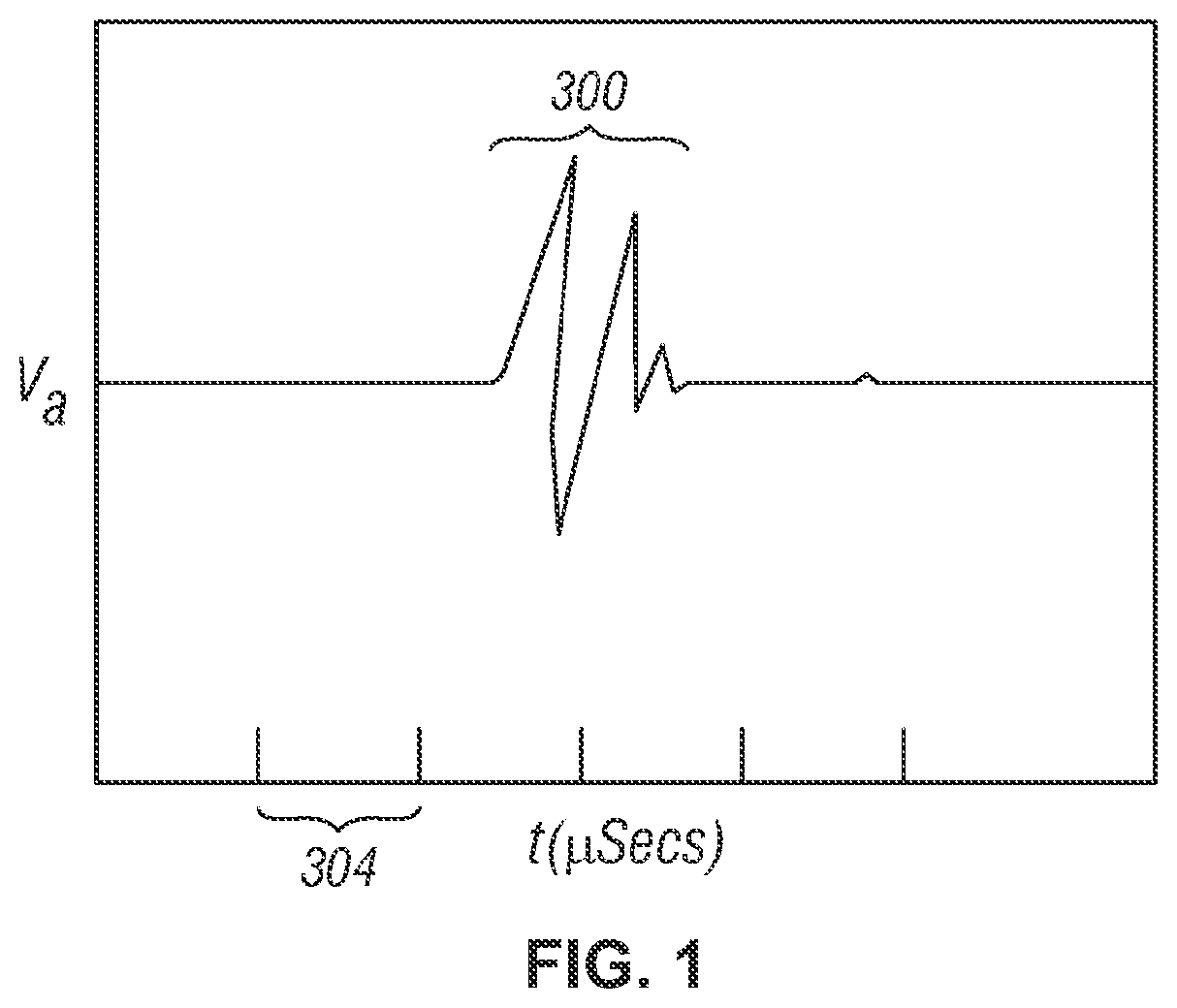
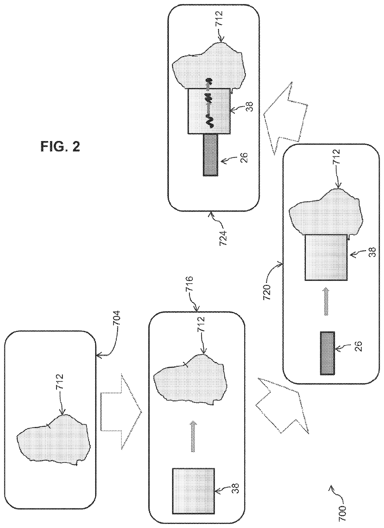
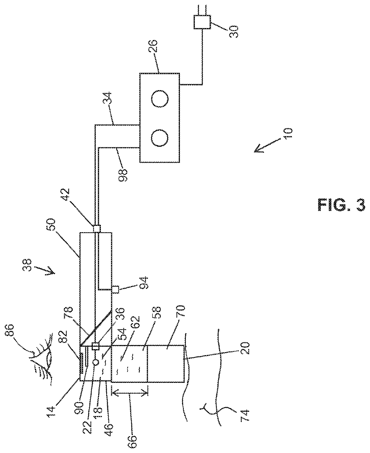
CROSS-REFERENCE TO RELATED APPLICATIONS[0001]This application is a national phase application under 35 U.S.C. 371 of International Application No. PCT / US2016 / 032069, filed May 12, 2016, which claims priority to U.S. Provisional Patent Application No. 62 / 160,147, filed May 12, 2015; and U.S. Provisional Patent Application No. 62 / 277,796, filed Jan. 12, 2016; all of which applications are incorporated by reference in their respective entireties.BACKGROUND1. Field of the Invention[0002]The present invention relates generally to methods of treatment for reducing adipose tissue using pressure waves. More particularly, but not by way of limitation, the present invention relates to methods of treatment for reducing subcutaneous adipose tissue using shockwaves.2. Description of Related ArtTreating Cellulite[0003]Excess body fat, localized adiposity, and cellulite represent important social problems. To date, techniques using radiofrequencies, ultrasound, and carbon dioxide have been studied...

Claims


Application Information

Patent Timeline
Patent Type & Authority Patents(United States)
IPC IPC(8): A61N7/00A61H23/00A61B17/225
CPCA61H23/008A61N7/00A61B17/225A61H2201/0153A61H2201/1207A61H2201/1238A61H2205/06A61H2205/083A61H2205/086A61H2205/108A61H2207/00A61N2007/0008A61N2007/0034A61N2007/0065
Inventor CAPELLI, CHRISTOPHER C.ROBERTSON, DAVID
Owner SOLITON
Learn More