Method for detecting t cell response to specific antigens in whole blood

a t cell response and specific antigen technology, applied in the field of detecting t cell response to specific antigens in whole blood, can solve the problems of lack of definitive information methodological, inability to define functional cell types, and little understanding of the development and homeostasis of ag-specific human memory/effector t cells in health or diseas

Inactive Publication Date: 2001-07-05
BECTON DICKINSON & CO +1
View PDF0 Cites 55 Cited by
  • Summary
  • Abstract
  • Description
  • Claims
  • Application Information

AI Technical Summary

Problems solved by technology

Despite widespread agreement that the basic unit of function of the T cell arm of the cellular immune system is the collection of memory / effector T cell clones responsive to a specific antigen(Ag), there is very little understanding of the development and homeostasis of Ag-specific human memory / effector T cells in either health or disease.
In large part, this lack of definitive information is methodological; i.e. the simple lack of tools to reliably quantitate and functionally evaluate Ag-specific T cells in the human.
New knowledge of immunological processes in the past decade, however, have highlighted serious problems with this approach.
While this addition to the measurement of proliferative responses represents a clear advance, in terms of a more comprehensive measure of T cell activation, these assays do not define the functional cell types that participate in these responses.
However, these assays are extremely laborious--especially if set up to measure the multiple, potentially relevant cytokines which may be produced by functionally heterogeneous Ag-specific memory / effector T cells--and therefore are not amenable to routine use in the clinical laboratory.
Moreover, the sensitivity of these assays may be limited by (1) conditions which do not mimic in vivo cellular environments which may be more clinically relevant, (2) T cell stimulation conditions that must necessarily compromise between conditions optimal for T cell activation and those compatible with the single cell detection strategy of the assay (e.g. limiting dilution conditions or spot counting on nitrocellulose covered culture wells), and (3) vulnerability to some loss of Ag-reactive cells from activation-induced apoptosis (as these assays require from 1-2 to many days of in vitro culture, and activation-induced apoptosis, as measured by DNA strand breaks, can initiate in as few as 12-19 hours).

Method used


Image

  • Method for detecting t cell response to specific antigens in whole blood
  • Method for detecting t cell response to specific antigens in whole blood
  • Method for detecting t cell response to specific antigens in whole blood

Examples


example 1

Cell Preparation and Antigen Stimulation

[0048] Heparinized whole blood samples were collected from normal or diseased donors using heparin Vacutainer.TM. blood collection tubes (Becton Dickinson VACUTAINER Systems, Franklin Lakes, N.J.). One ml aliquots of whole blood were dispensed into 16.times.125 polypropylene tissue culture tubes (Corning Costar Corporation, Cambridge, Mass.).

[0049] Appropriately titred specific or control Ag preparations (60 .mu.l / ml) and in most instances (except as noted), 4 .mu.g anti-CD28 MAbs were added to 1 ml whole blood aliquots. Cultures were incubated at a 5.degree. slant at 37.degree. C. in a humidified 5% CO2 atmosphere for 1 hour with slight agitation to improve APC / T cell interaction, and an additional final 5 hours including a final concentration of 10 .mu.g / ml of Brefeldin A (a relatively non-toxic, but potent, inhibitor of intracellular transport which prevents secretion of any produced cytokines. 20 mM Na2EDTA for a final concentration of 2 m...

example 2

Immunofluorescent Staining

[0050] Cell preparations frozen as described above were rapidly thawed in a 37.degree. C. water bath and then washed once with cold dPBS prior to resuspension in fixation / permeabilization solution (Becton Dickinson Immunocytometry Systems, San Jose, Calif.) at 1.times.10.sup.6 cells / 500 ul, and incubation for 10 minutes at room temperature in the dark. These cells were washed twice with dPBS with BSA and sodium azide, and then were stained protected from light with directly conjugated MAbs for 30 minutes. In some experiments, the freezing step was omitted, and cells freshly harvested after Ag activation were cell surface stained first, and then fixed / permeabilized / washed, and then stained for intracytoplasmic Ags. After staining, the cells were washed, refixed in 1% paraformaldehyde in dPBS, and then kept protected from light at 4.degree. C. until analysis on the flow cytometer.

example 3

Flow Cytometric Analysis

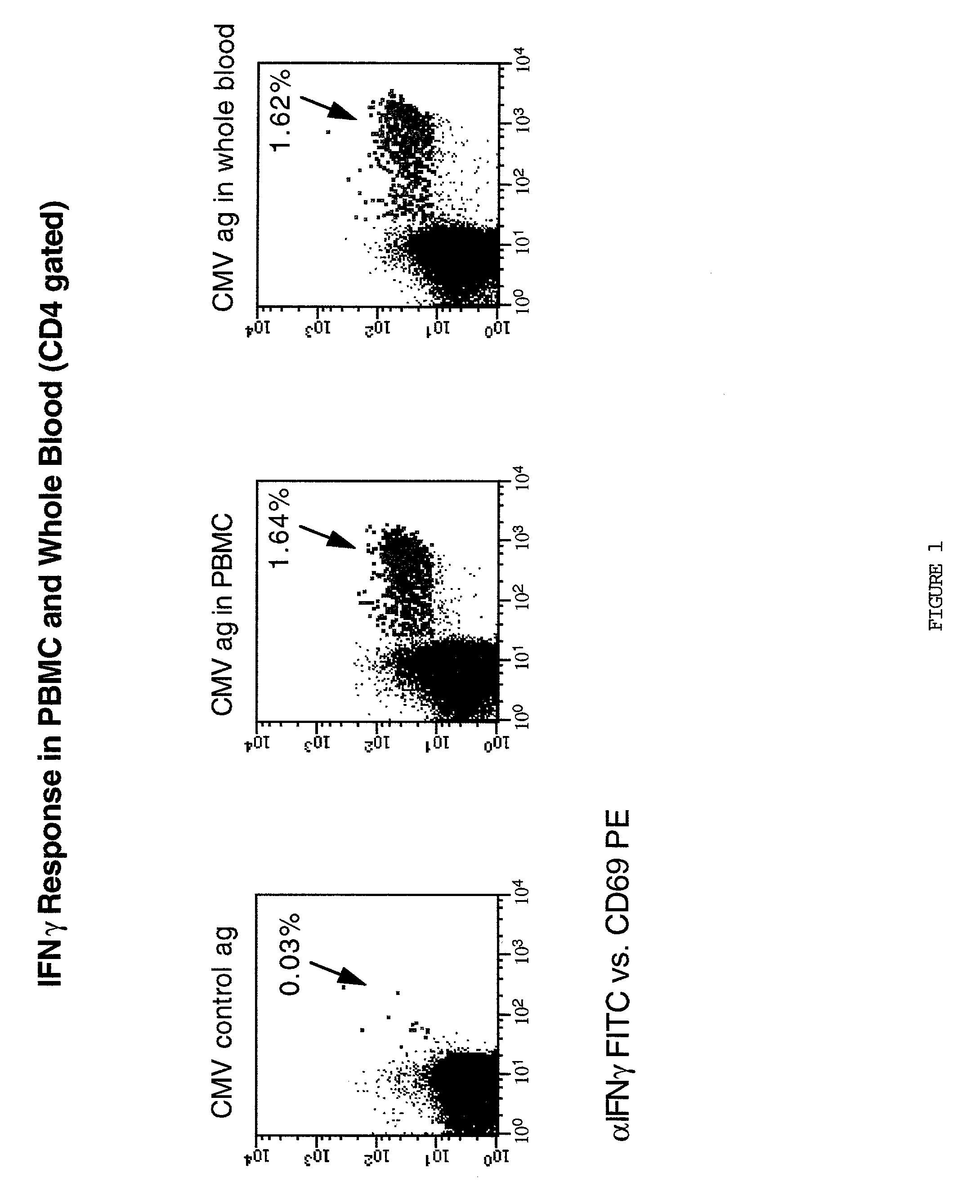
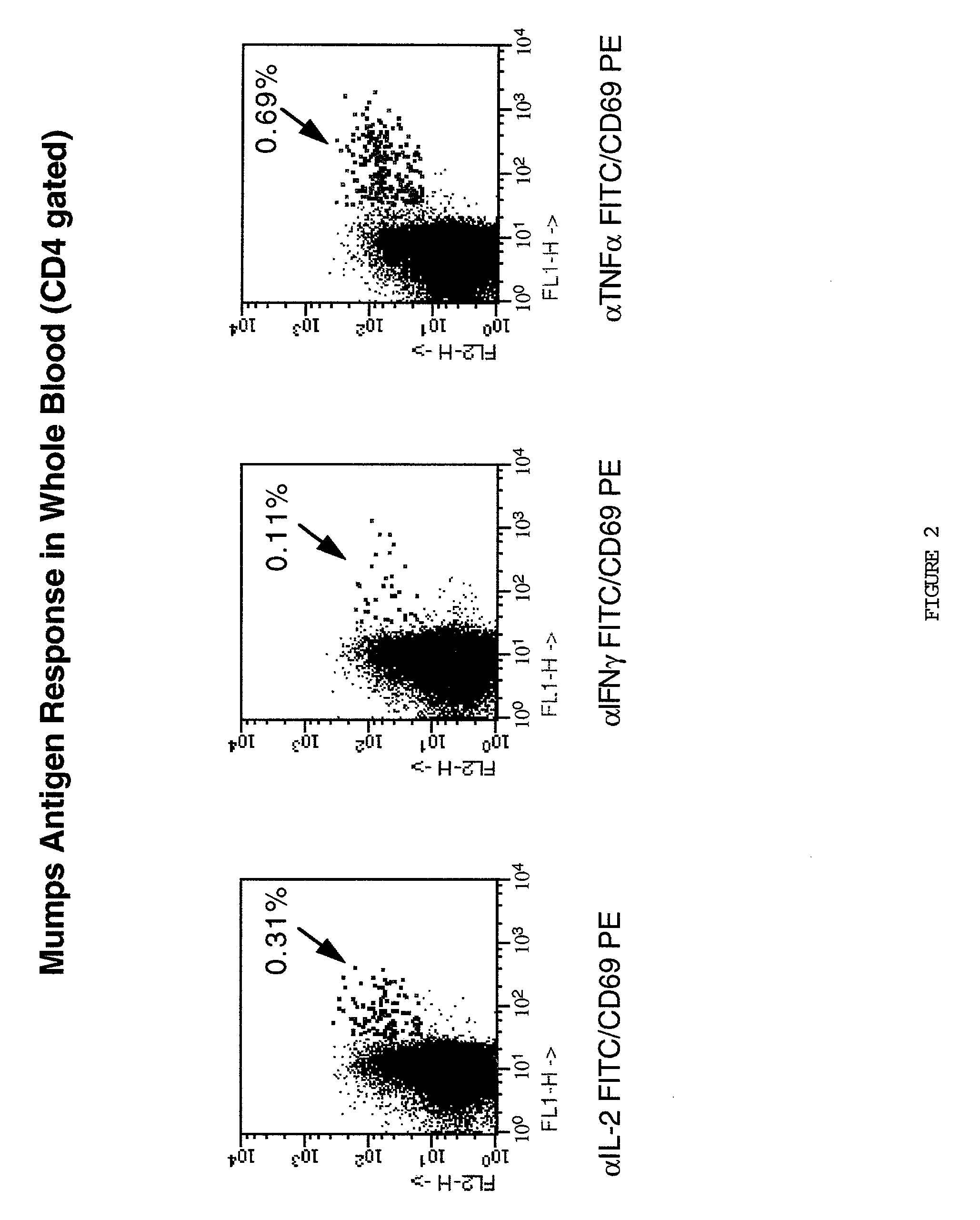
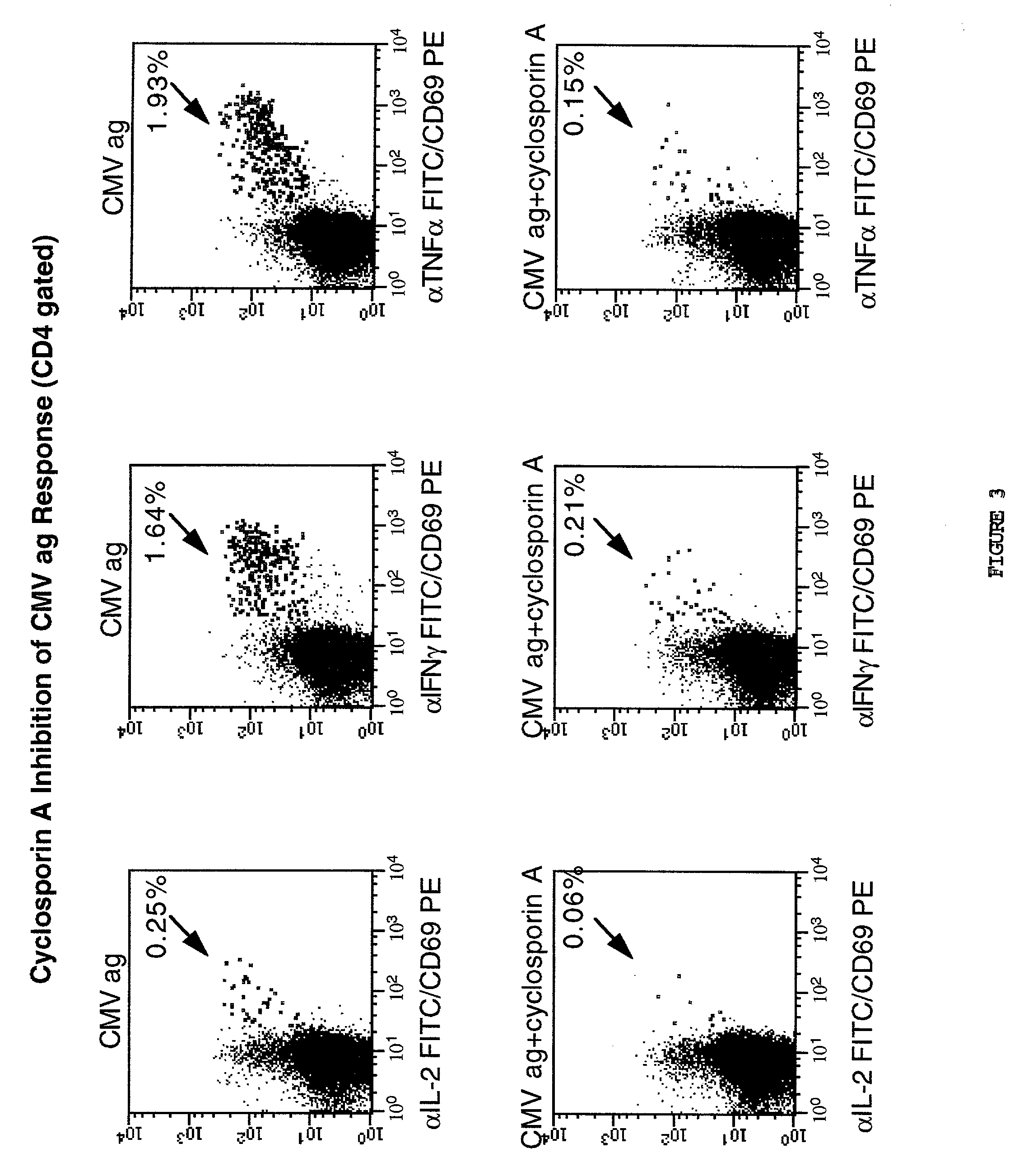
[0051] Up to six parameter analysis was performed on a modified FACSort.TM. (Becton Dickinson) flow cytometer equipped with a second 532 nm line diode laser (BDIS) using FITC, phycoerythrin(PE), and peridinin chlorophyll protein (PerCP), and allophycocyanin (APC) as the 4 fluorescent parameters, using methods of cytometer set up and data acquisition which are well known in the art. Otherwise 5 parameter analysis using up to three fluorescent parameters was conducted on a standard FACScan.TM. flow cytometer. For a typical analysis, 50,000 events were acquired, gated on CD4 expression and a light scatter gate designed to include only viable lymphocytes (most files required "fine-tuning" of gating during analysis on these same parameters, leaving 48,000 events for the final profiles). In some analyses, additional "live" gating based on a fluorescent parameter (for example, CD69 reactivity) was performed to enhance the sampling of small populations. List mode mul...


PUM

PropertyMeasurementUnit
concentrationaaaaaaaaaa
frequencyaaaaaaaaaa
adhesionaaaaaaaaaa

Abstract

This invention comprises a novel approach to the assessment of antigen-specific T cells that quantitates and characterizes these cells with unprecedented clarity, and importantly, because it is performed in whole blood, is amenable to routine use in the clinical immunology laboratory. The methodology offers an improved flow cytometric intracellular cytokine assay in whole blood that can simultaneously measure multiple T cell subsets expressing multiple cytokines from a single whole blood culture. Evaluation of whole blood antigen specific cytokine responses has the important advantage of assessing T cell activation in the presence of ALL types of MHC autologous antigen presenting cells present in the native sample. It also has the advantage of enabling a culture system (whole blood) which can reflect effects of systemic environments (i.e. drug augmentation or suppression) on T cell responses to specific stimuli including antigen, by either culturing in the presence of such drug or analyzing the blood of a human or animal receiving such drug.

Description

[0001] This application is a continuation-in-part of copending application Ser. No. 08 / 760,447 filed Dec. 6, 1996.BACKGROUND OF INVENTION[0002] Despite widespread agreement that the basic unit of function of the T cell arm of the cellular immune system is the collection of memory / effector T cell clones responsive to a specific antigen(Ag), there is very little understanding of the development and homeostasis of Ag-specific human memory / effector T cells in either health or disease. In large part, this lack of definitive information is methodological; i.e. the simple lack of tools to reliably quantitate and functionally evaluate Ag-specific T cells in the human. For many years, assays of antigen specific T cell function relied on proliferation of cultured PBMC as the measure of the responding T cells.[0003] New knowledge of immunological processes in the past decade, however, have highlighted serious problems with this approach. Proliferative responses require days to manifest, and it...

Claims


Application Information

Patent Timeline
Patent Type & Authority Applications(United States)
IPC IPC(8): G01N33/569
CPCG01N33/56972
Inventor MAINO, VERNON C.SUNI, MARIA
Owner BECTON DICKINSON & CO
Why Eureka
Try Eureka