
Cell-culture and polymer constructs

a cell culture and polymer technology, applied in the field of cell culture and polymer constructs, can solve the problems of insufficient cell culture, insufficient cell culture, and insufficient cell culture, and achieve the effect of efficient cell propagation in vitro

Inactive Publication Date: 2002-09-05
HUNGERFORD DAVID S +4
View PDF16 Cites 17 Cited by
  • Summary
  • Abstract
  • Description
  • Claims
  • Application Information

AI Technical Summary

Benefits of technology

[0230] The herein disclosed invention seeks to perfect cartilage repair surgery. The invention envisions overcoming many of the failings practiced by the medical profession and, particularly, the commercial practice of cartilage repair. The invention seeks to improve cartilage repair surgery in the following ways:[0231] 1) The invention will make available sufficient quantities of compatible chondorcytes required for repair (and in a relatively short period of time) by growing or culturing the chondrocytes in a spinning culture chamber and, preferably, at reduced oxygen levels.[0232] 2) The patient's own chondrocytes will be used to culture cells for plating onto the scaffold. Cadaver cells will not have to be used.[0233] 3) The invention envisions rapidly growing chondrocytes in culture.[0234] 4) Chondrocytes cultured will be characterized as having an approximately normal phenotype.[0235] 5) Scaffolds prepared for transplant will be physiologically compatible and resorbable into the body.[0236] 6) Cell culture and surgical facilities will be at a single location which will assume rapid and efficient surgical procedures envisioning a high surgical success rate.[0237] This should be contrasted with the correct medical procedures, which may be outlined as follows:[0238] Taking chondrocytes from the patients for subsequent growing in a chamber (non-spinning and at ordinary oxygen levels)which requires a relatively long time period to grow sufficient quantities of the chondorcytes for subsequent transplanting into patient.[0239] Taking chondrocytes from a cadaver, growing the chondrocytes and plating on a scaffold (or other plastic or inorganic support) and thereafter transplanting into the patient.[0240] Obviously, many modifications may be without departing from the basic spirit of the present invention. Accordingly, it will be appreciated by those skilled in the art that within the scope of the appended claims, the invention may be practiced other than has been specifically described herein.

Problems solved by technology

Articular cartilage is frequently damaged in the course of common activities.
In addition, osteoarthritis leads to the initial focal wear of articular cartilage in many joints, particularly the hip and the knee.
Alternative concepts are in development, but none are approved.
No current therapy other than cadaver cartilage transplant offers this potential.
Cadaver cartilage transplant is ineffective over the long term, is expensive and transplant material is not readily available.
In both instances, success is dependent on an uncontrolled response from the body in an uncontrolled environment.
In the past, such response has been unpredictable and the development of such a response has required long periods of inactivity on the part of the patient.
These methods are not able to provide sufficient numbers of chondrocytes with unaltered phenotype needed for analysis.
However, the ease of recovering high yields of viable and functional cells from microcarriers has been a problem.
The most common method of cell harvesting used is by trypsinizaion, which may not completely retrieve cells from the microcarriers.
In addition, the patentees found that harvesting cells from collagen-only beads presented difficulties in separation of the cells from the media in that there was contamination of the cells by soluble fragments of collagen.

Method used


Image

  • Cell-culture and polymer constructs
  • Cell-culture and polymer constructs
  • Cell-culture and polymer constructs

Examples


Embodiment Construction

[0140] We have discovered that human chondrocytes and other cells types such as osteoblastic cells can be successfully incorporated into chitosan constructs. The cells remain viable and are metabolically active indicated by their continued protein synthesis. They do not proliferate but continue to produce tissue specific matrix components. Chondrocytes produce collagen type II whereas osteoblasts produce collagen type I. The section below describes the (1) examples of cells that can be used in chitosan constructs, (2) types of chitosan tested in our laboratory, and (3) method of preparing the construct.

DESCRIPTION AND OPERATION

[0141] Differentiated chondrocytes or undifferentiated precursor cells from the bone marrow, periosteum and perichondrium can be incorporated into the chitosan scaffold described below. Several types of chitosan have been tested and characterized in our laboratory as shown in Table I Below. These chitosans were purchased from different sources.

5TABLE IV Commer...


PUM

PropertyMeasurementUnit
volume capacityaaaaaaaaaa
surface areaaaaaaaaaaa
volume capacityaaaaaaaaaa

Abstract

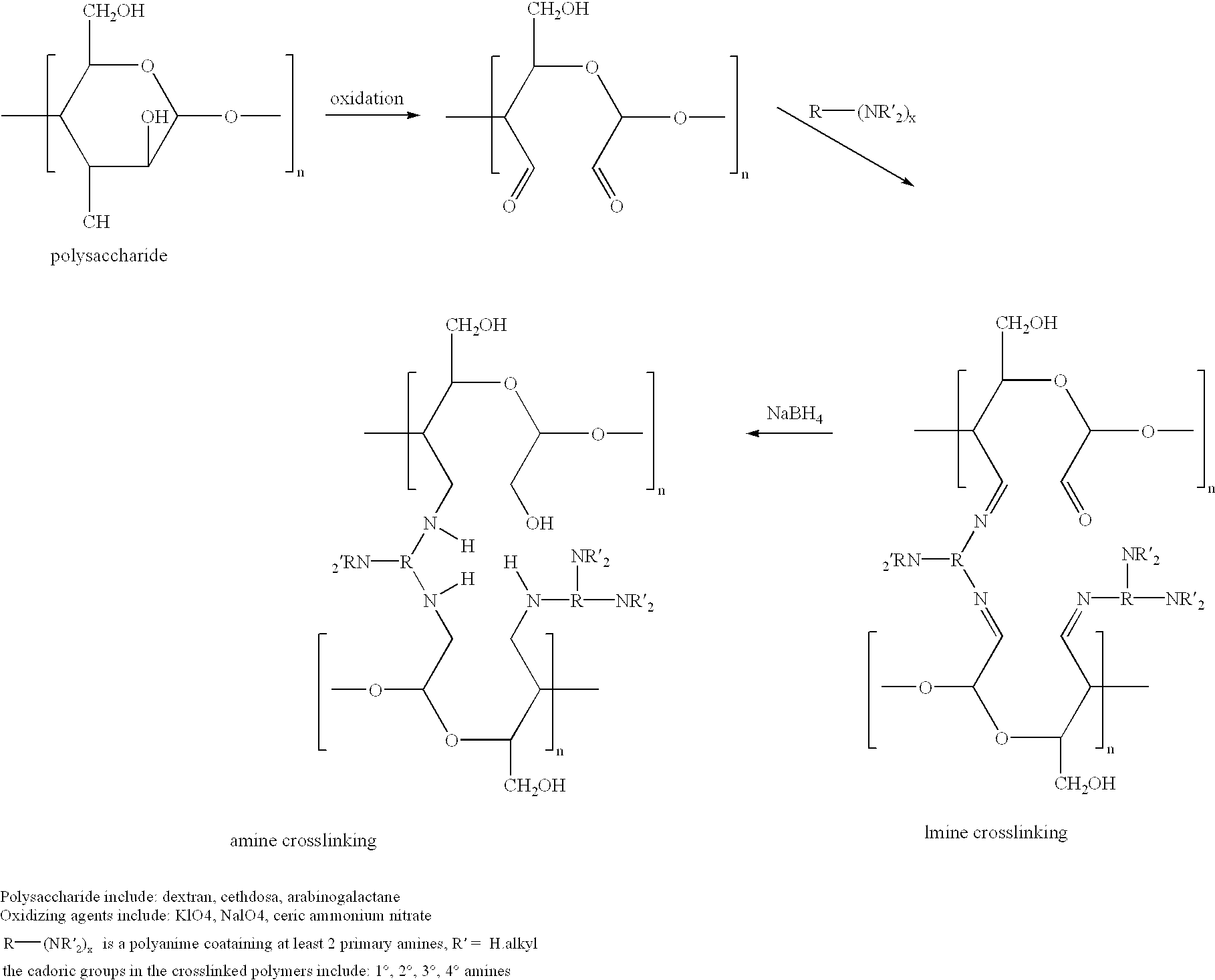
Cells grown on a microcarrier are separated from the microcarrier by enzymatically digesting the microcarrier. More specifically, chondrocytes may be grown on dextran microcarrier beadlets and then the beadlets digested using dextranase to separate the chondrocytes from the carrier. Cells can also be grown on chitosan microcarriers to be used for implantation. In addition, cells can be grown on polysaccharide polymers to be used as implant devices. Various polymers serve as scaffolds for cells to be used for implantation. The polymers can be used for cell culture as well as for preparing scaffolds useful for tissue replacement such as cartilage tissue.

Description

[0001] This application is related to provisional applications Serial No. 60 / 081,016 filed Apr. 8, 1998 and Serial No. 60 / 104,842 filed Oct. 20, 1998.[0002] The herein disclosed invention finds applicability in the field of cell culture, as well as in the field of tissue substitute for tissue replacement.[0003] Attempts at replacing or rebuilding diseased or damaged structures in the human body go back to 3000 B.C. It was not until the middle of the 1900's, however, that the use of synthetic materials for rebuilding body structures met with widespread and reproducible success. Advances in material science and biomaterials and science have afforded much of the success. The need for new and better implants exists in every field of medicine in which disease or trauma can be treated surgically.[0004] As technology advances continue to improve the state of the art, the standards for successful implants continue to improve including strength, biocompatibility and elasticity. The new resea...

Claims


Application Information

Patent Timeline
Patent Type & Authority Applications(United States)
IPC IPC(8): A61F2/08A61K9/16A61K9/50A61K35/12A61K47/36A61L27/38C08B15/00C08B33/00C08B37/00C08B37/02C08H1/00C08J3/12C12N5/00C12N5/077
CPCA61F2/08A61F2/30756A61K9/1652A61K9/5036A61K35/12A61K47/36A61L27/3817A61L27/3852A61L27/3895C08B15/005C08B33/00C08B37/00C08B37/0021C08B37/006C08H1/00C08J3/12C08J2305/08C12N5/0075C12N5/0654C12N5/0655C12N2500/02C12N2531/00C12N2533/70C12N2533/72A61L2430/06
Inventor HUNGERFORD, DAVID S.FRONDOZA, CARMELITA G.SOHRABI, AFSHINSHIKANI, ALAN H.DOMB, ABRAHAM J.
Owner HUNGERFORD DAVID S
Learn More