In situ langerhans cell vaccine

a technology of in situ langerhans cells and vaccines, which is applied in the field of in situ langerhans cell vaccines, can solve the problems of low number of dendritic cells, unable to obtain dendritic cells in sufficient quantities, and unable to meet the requirements of francotte or urbain

Inactive Publication Date: 2002-09-19
US SOUTHWESTERN MEDICAL CENT
View PDF6 Cites 33 Cited by
  • Summary
  • Abstract
  • Description
  • Claims
  • Application Information

AI Technical Summary

Problems solved by technology

The efficacy of DCs in delivering antigens in such a way that a strong immune response ensues i.e., "immunogenicity", is widely acknowledged, but the use of these cells for delivering antigens is hampered by the fact that there are very few DCs in any given organ.
While DCs can process foreign antigens into peptides that must be recognized by immunologically active T cells (i.e., dendritic cells accomplish the phenomenon of "antigen presentation"), the low numbers of dendritic cells prohibits their use in identifying immunogenic peptides.
However Francotte, Urbain and patent application WO 91 / 13632 do not provide a practical method of using dendritic cells as an adjuvant to activate the immune response, because both of these methods depend on ex vivo techniques wherein dendritic cells are obtained from spleen, an impractical source of cells for most therapies or immunization procedures.
In addition, neither Francotte nor Urbain provides a method to obtain dendritic cells in sufficient quantities to be clinically useful.
While these methods have had modest success, tolerance has not been achieved.
(TGF-.beta.) using an adenoviral vector prevents the reduction of DCs generally seen with adenovirus infection and also increases the numbers and prolongs the survival of the infected DCs in the spleen of a host to whom the DCs are administered.
While modification of DCs may be an attractive approach to the therapy of cancer, infectious disease, foreign graft rejection, allergic disorders, GVH disease and autoimmune disorders, there are potential problems associated with such an approach.
Although many DC-based vaccine protocols have been developed, the processes for developing them are time consuming and costly because they require ex vivo DC manipulation including isolation and expansion of DCs from individual hosts, manipulation of the maturational state of the DCs in culture, loading of the DCs in culture and administration of the loaded DCs back into the host.
Such cumbersome, time-consuming and costly approaches limit the clinical application of DC vaccines.
One potential problem associated with delivering GM-CSF fused to TAA is that GM-CSF binds to other cell types which may lead to tolerance rather than immunity.
However, if chemokine genes are injected directly without any pulsing with a particular antigen, fine antigen specificity may be lost which could lead to an autoimmune response.
This method is limited because penetration of molecules into the skin is not very efficient.
In addition, it is very difficult to apply large molecules through the skin and therefore, the method is likely only effective with small peptides, as indicated (preferably the antigen is a peptide of 2 to 20 amino acids).

Method used


Image

  • In situ langerhans cell vaccine
  • In situ langerhans cell vaccine
  • In situ langerhans cell vaccine

Examples


examples

[0074] Example 1: Preparation of EVA polymer rods

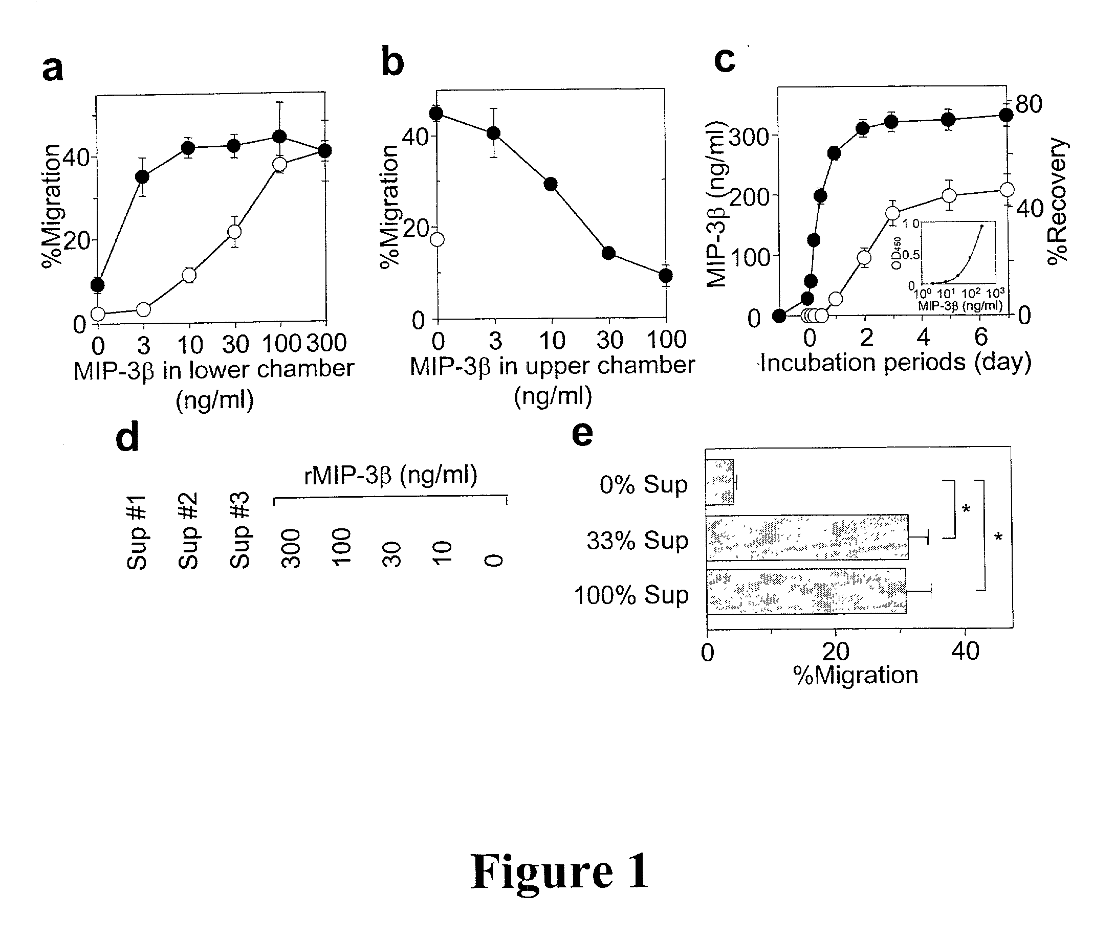
[0075] All polymer rods were prepared in the Brown University laboratory of Dr. Robert F. Valentini. MIP-3.beta. (900 ng / rod) was incorporated into the EVA polymer rods as described by Kim & Valentini, 1997, Biomaterials 18:1175-1184. Briefly, lyophilized MIP-3.beta. was reconstituted in PBS, mixed with 1% BSA (1.8 mg / rod added as a carrier protein), snap frozen in liquid nitrogen, and re-lyophilized. EVA copolymer pellets (DuPont, Wilmington, Del.) were washed extensively and dissolved in methylene chloride to obtain a 1% solution (w / v). The lyophilized MIP-3.beta.-BSA mixture was then added to the polymer solution with the ratio of 40:60 by weight, vortexed, snap frozen, and re-lyophilized. The resulting powder was melt-extruded at 50.degree. C. into continuous polymer rods with a diameter of 0.7 mm. Rods were cut into 10 mm pieces and coated by dipping in a 5% EVA-methylene chloride solution. EVA rods containing BSA or OVA (1.8 mg / ...

example 4

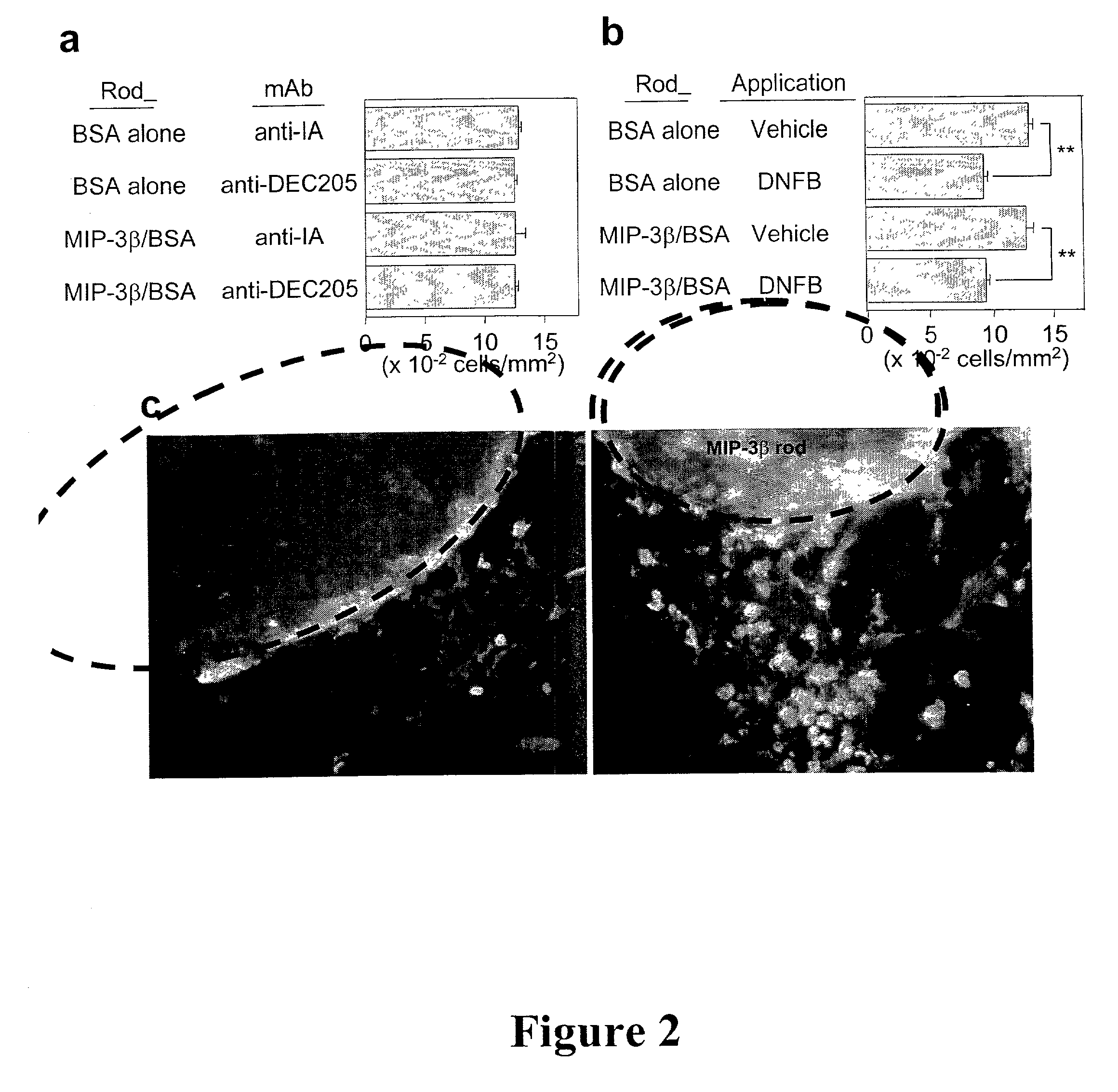
[0085] LC entrapment

[0086] After 24 hr pre-incubation in PBS at 37.degree. C., a MIP-3.beta. or BSA rod was cut into 4 short pieces (2.5 mm length) and implanted subcutaneously into a mouse with 200 .mu.l of PBS using a 20G1 / 2 needle. At 24 hr post-implantation, 20 .mu.l of 0.5% dinitroflourobenzene (DNFB) or 3% fluorescein isothiocyanate (FITC) was carefully applied over the implantation sites (10 mm-diameter circles marked with black ink immediately after implantation).

[0087] Subcutaneous implantation of EVA polymer rods containing MIP-3.beta. (900 ng / rod / animal) did not affect the number of LC that remained in the overlaying epidermis (FIG. 2a) or cause significant accumulation of IA.sup.+ cells (IA.sup.+ serves as a marker for APCs) around the implanted rods at 24 hour pont-implantation. In an attempt to induce LC maturation and elevate MIP-3.beta. responsiveness, DNFB was applied over the implantation sites. Significant (25-30%) reduction was observed in surface LC densities 2...

example 6

[0092] Loading of LC in situ

[0093] For in situ LC loading with antigen, a pre-soaked OVA rod (OVA was used as a model tumor associated antigen) containing 1.8 mg / rod / animal was cut into 4 pieces and co-implanted with a MIP-3.beta. or BSA rod into an animal (C57BL / 6 mice) and the implantation site was painted with DNFB 24 hr later. FIGS. 4a and 4b show induction of tumor-specific CTL activities an protective immunity by in situ LC loaded cells in three groups. The first group of C57BL / 6 mice received co-implantation of MIP-3.beta. rods+OVA rods on the abdomen followed by application of DNFB at the implantation site (circles), the second group received coimplantation of BSA rods+OVA rods on the abdomen followed by application of DNFB at the implantation site (squares) and the third group received implantation of MIP-3.beta. rods on the back and OVA rods on the abdomen, followed by DNFB application at the site of implantation of the MIP-3.beta. rods on the back. Spleen cells were harv...


PUM

PropertyMeasurementUnit
concentrationsaaaaaaaaaa
lengthaaaaaaaaaa
diameteraaaaaaaaaa

Abstract

A method for entrapping migratory antigen presenting cells (APCs) and particularly Langerhans cells (LCs,) in vivo is provided. The method entails creating an artificial gradient of APC-attracting chemotactic factor in the homing path of APCs in vivo. Also provided is a composition for entrapping APCs and particularly, migratory LCs. In addition, a method for loading APCs in situ with antigen is provided. The method comprises entrapping APCs in vivo and subsequently loading the APCs in situ with antigen. Correspondingly, a composition for loading APCs in situ is also provided. Further provided is a method for stimulating the migration of entrapped APCs to draining lymph nodes. The ability to stimulate the migration of entrapped APCs to draining lymph nodes is useful, inter alia, for regulating an immune response in a subject. In addition, an in situ APC-based vaccine is provided which does not require any time-consuming, costly ex vivo manipulations.

Description

[0002] The present invention relates to a method for regulating an immune response in a mammalian subject wherein an artificial gradient of a chemotactic factor is created in vivo and wherein said gradient allows for the transient entrapment of antigen presenting cells (APCs), such as Langerhans cells (LCs). The transient entrapment of APCs promotes the loading of the entrapped APCs with one or more immunoregulatory molecules, e.g. one or more antigens, one or more immunostimulatory molecules and / or one or more immunosuppressive molecules. After loading, the transiently entrapped APCs migrate to draining lymph nodes (DLN). The method of the present invention is useful for regulating an immune response in a mammalian subject, e.g., for treating or preventing cancer, treating or preventing infectious disease, treating or preventing autoimmune disease, treating or preventing allergic disorders, prolonging graft survival, treating or preventing graft versus host (GVH) disease and for in...

Claims


Application Information

Patent Timeline
Patent Type & Authority Applications(United States)
IPC IPC(8): A61K9/00A61K38/19A61K38/22A61K38/34A61K39/00
CPCA61K9/0092A61K39/00A61K39/0011A61K2039/5154C12N2501/21C12N2501/23A61K38/195A61K39/001106A61K39/001111A61K39/001151A61K39/001156A61K39/001157A61K39/001163A61K39/001164A61K39/00117A61K39/001182A61K39/001184A61K39/001186A61K39/001191A61K39/001192A61K39/001195
Inventor TAKASHIMA, AKIRAKUMAMOTO, TADASHI
Owner US SOUTHWESTERN MEDICAL CENT
Why Eureka
Try Eureka