Purified hepatitis C virus envelope proteins for diagnostic and therapeutic use

a technology of hepatitis c virus and envelope proteins, which is applied in the direction of viruses, peptides/protein ingredients, fungi, etc., can solve the problems of much more expensive manufacturing of hbsag from mammalian cells compared with yeast-derived hepatitis b vaccines

Inactive Publication Date: 2003-02-20
INNOGENETICS NV
View PDF2 Cites 4 Cited by
  • Summary
  • Abstract
  • Description
  • Claims
  • Application Information

AI Technical Summary

Problems solved by technology

In the case of hepatitis B for example, manufacturing of HBsAg from mammalian cells was much more costly compared with yeast-derived hepatitis B vaccines.

Method used


Image

  • Purified hepatitis C virus envelope proteins for diagnostic and therapeutic use
  • Purified hepatitis C virus envelope proteins for diagnostic and therapeutic use
  • Purified hepatitis C virus envelope proteins for diagnostic and therapeutic use

Examples


example 2

Construction of HCV Recombinant Plasmids

[0306] 2.1. Constructs Encoding Different Forms of the E1 Protein

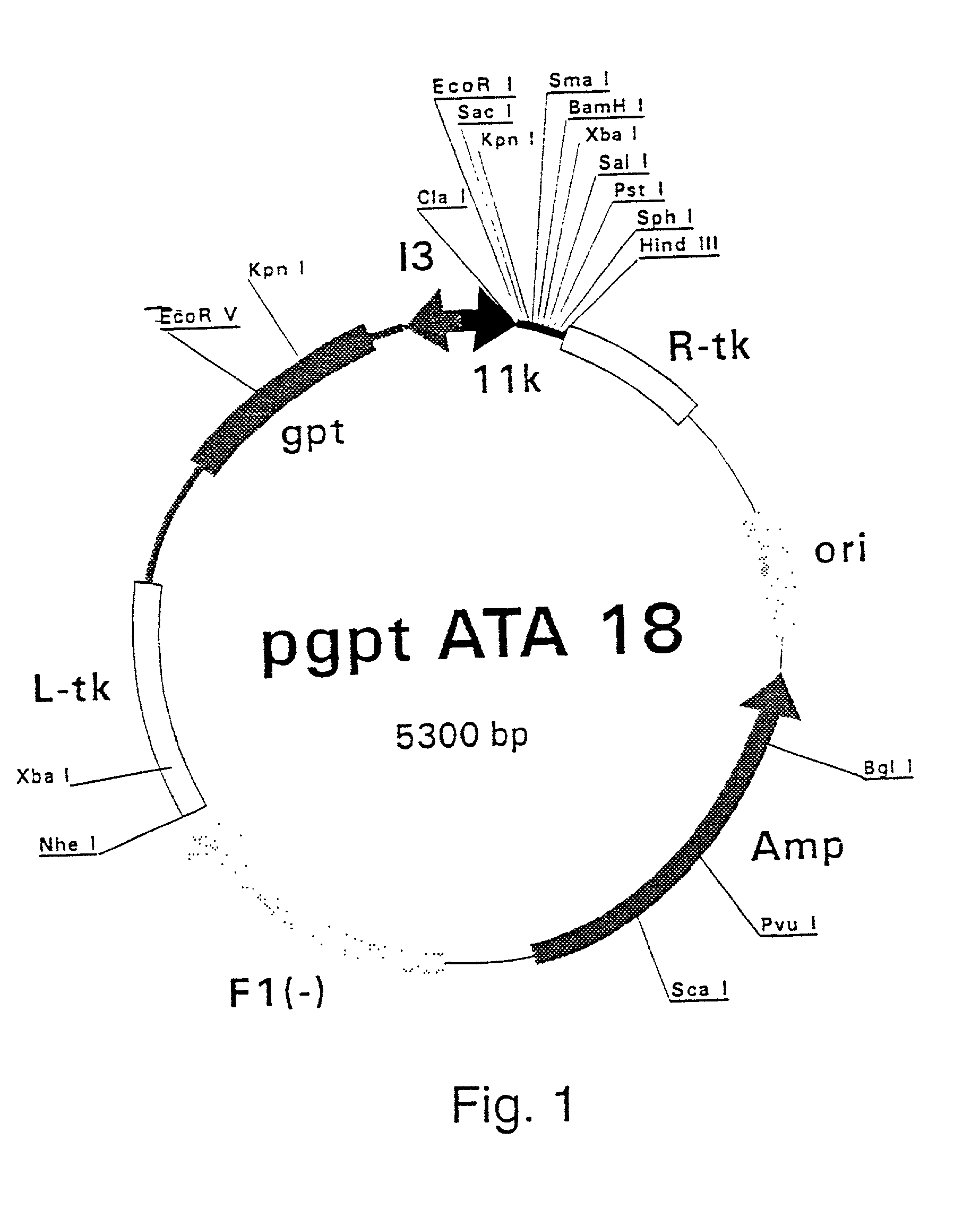
[0307] Polymerase Chain Reaction (PCR) products were derived from the serum samples by RNA preparation and subsequent reverse-transcription and PCR as described previously (Stuyver et al., 1993b). Table 1 shows the features of the respective clones and the primers used for amplification. The PCR fragments were cloned into the Sma I-cut pSP72 (Promega) plasmids. The following clones were selected for insertion into vaccinia recombination vectors: HCCl9A (SEQ ID NO 3), HCCl10A (SEQ ID NO 5), HCCl11A (SEQ ID NO 7), HCCl12A (SE: ID NO 9), HCCl13A (SEQ ID NO 11) and HCCl17A (SEQ ID NO 13) as depicted in FIG. 21. cDNA fragments containing the E1-coding regions were cleaved by EcoRI and HindIII restriction from the respective pSP72 plasmids and inserted into the EcoRI / HindIII-cut pgptA-18 vaccinia recombination vector (described in example 1), downstream of the 11K vaccinia virus late p...

example 3

Infection of Cells with Recombinant Vaccinia Viruses

[0317] A confluent monolayer of RK13 calls was infected at a m.o.i. of 3 with the recombinant HCV-vaccinia viruses as described in example 2. For infection, the cell monolayer was washed twice with phosphate-buffered saline pH 7.4 (PBS) and the recombinant vaccinia virus stock was diluted in MEM medium. Two hundred .mu.l of the virus solution was added per 10.sup.6 cells such that the m.o.i. was 3, and incubated for 45 min at 24.degree. C. The virus solution was aspirated and 2 ml of complete growth medium (see example 2) was added per 10.sup.6 cells. The cells were incubated for 24 hr at 37.degree. C. during which expression of the HCV proteins took place.

example 4

Analysis of Recombinant Proteins by Means of Western Blotting

[0318] The infected cells were washed two times with PBS, directly lysed with lysis buffer (50 mM Tris.HCl pH 7.5, 150 mM NaCl, 1% Triton X-100, 5 mM MgCl.sub.2, 1 .mu.g / ml aprotinin (Sigma, Bornem, Belgium)) or detached from the flasks by incubation in 50 mM Tris.HCL pH 7.5 / 10 mM EDTA / 150 mM NaCl for 5 min, and collected by centrifugation (5 min at 1000 g). The cell pellet was then resuspended in 200 .mu.l lysis buffer (50 mM Tris.HCL pH 8.0, 2 mM EDTA, 150 mM NaCl, 5 mM MgCl.sub.2 aprotinin, 1% Triton X-100) per 10.sup.5 cells. The call lysates were cleared for 5 min at 14.000 rpm in an Eppendorf centrifuge to remove the insoluble debris. Proteins of 20 .mu.l lysate were separated by means of sodium dodecyl sulphate-polyacrylamide gel electrophoresis (SDS-PAGE). The proteins were then electro-transferred from the gel to a nitrocellulose street (Amersham) using a Hoefer HSI transfer unit cooled to 4.degree. C. for 2 hr at...


PUM

PropertyMeasurementUnit
molecular weightaaaaaaaaaa
pHaaaaaaaaaa

Abstract

The present invention relates to a method for purifying recombinant HCV single or specific oligomeric envelope proteins selected from the group consisting of E1 and / or E1 / E2 characterized in that upon lysing the transformed host cells to isolate the recombinantly expressed protein a disulphide bond cleavage or reduction step is carried out with a disulphide bond cleavage agent. The present invention also relates to a composition isolated by such a method. The present invention also relates to the diagnostic and therapeutic application of these compositions. Furthermore, the invention relates to the use of HCV E1 protein and peptides for prognosing and monitoring the clinical effectiveness and / or clinical outcome of HCV treatment.

Description

[0001] The present invention relates to the general fields of recombinant protein expression, purification of recombinant proteins, synthetic peptides, diagnosis of HCV infection, prophylactic treatment against HCV infection and to the prognosis / monitoring of the clinical efficiency of treatment of an individual with chronic hepatitis, or the prognosis / monitoring of natural disease.[0002] More particularly, the present invention relates to purification methods for hepatitis C virus envelope proteins, the use in diagnosis, prophylaxis or therapy of HCV envelope proteins purified according to the methods described in the present invention, the use of single or specific oligomeric E1 and / or E2 and / or E1 / E2 envelope proteins in assays for monitoring disease, and / or diagnosis of disease, and / or treatment of disease. The invention also relates to epitopes of the E1 and / or E2 envelope proteins and monoclonal antibodies thereto, as well their use in diagnosis, prophylaxis or treatment.[0003...

Claims


Application Information

Patent Timeline
Patent Type & Authority Applications(United States)
IPC IPC(8): A61K38/00A61K39/00A61K39/29G01N33/53A61K39/395A61P1/16A61P31/12C07K14/18C07K16/10C12N1/15C12N1/19C12N1/21C12N5/10C12N15/02C12N15/09C12N15/40C12P21/00C12P21/02C12P21/08C12Q1/70C12R1/865C12R1/91C12R1/92G01N33/576G01N33/577
CPCA61K39/00A61K39/29C07K14/005C07K16/109C07K2317/34C12N2770/24222C12N2710/24143C12N2770/24243Y10S435/81Y10S435/975Y10S435/803Y10S435/915A61K2039/5256C12N2770/24234A61K39/12A61P1/16A61P31/12
Inventor MAERTENS, GEERTBOSMAN, FONSDE MARTYNOFF, GUYBUYSE, MARIE-ANGE
Owner INNOGENETICS NV
Why Eureka
Try Eureka