Assay procedure using fluorogenic tracers
- Summary
- Abstract
- Description
- Claims
- Application Information
AI Technical Summary
Benefits of technology
Problems solved by technology
Method used
Image
Examples
example i
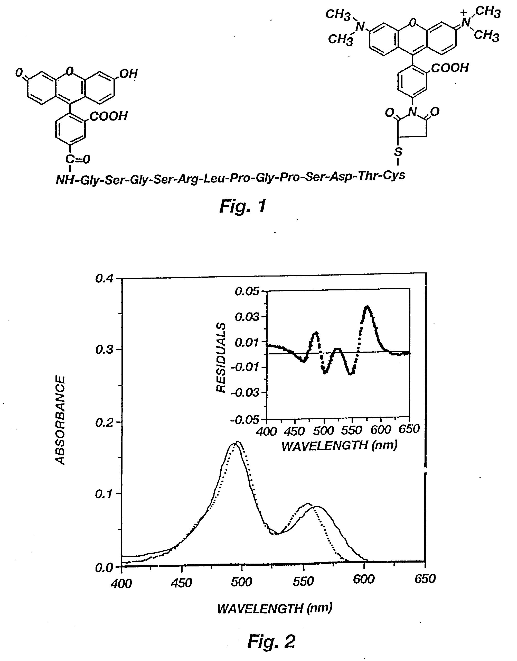
[0066] The hCG epitope peptide and its conjugate with dyes. A peptide from human chorionic gonadotrophin (hCG) was used as the spacer between fluorescein (F) and tetramethylrhodamine (T). Depicted in FIG. 1 is the structure of a hCG epitope peptide labeled with fluorescein and TMR. The peptide was identified by screening 221 (24) overlapping octapeptides synthesized on the tips of 96-pin solid supports as described in H. M. Geysen et al., Molecular Immunology, 23:709-715 (1986).
[0067] According to the Geysen method, a series of n-8 overlapping octapeptides were synthesized on the tips of 96-pin solid supports and tested for specific binding with anti-hCG using a ELISA procedure, where n is the number of amino acid residues in a sequence. A total number of 221 octapeptides was screened because hCG has two chains and a total number of 237 amino acid residues: R. B. Carlsen et al., J. Biol. Chem., 248:6810-6827 (1973); R. Bellisario et al., J. Biol. Chem., 248:6796-6809 (1973).
[0068] A...
example ii
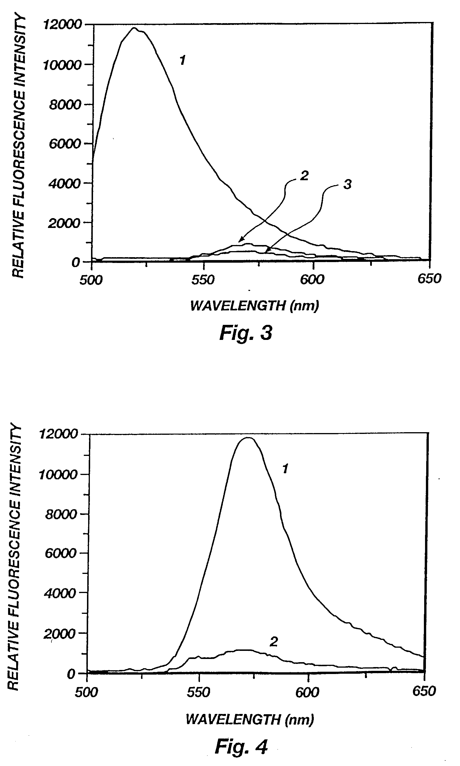
[0078] Binding of FpepT with anti-hCG Mab. The fluorescence spectra of FpepT upon binding to the anti-hCG Mab is presented in FIG. 5. The fluorescence of rhodamine (lambda max=570 nm) increased up to 5 fold with increasing antibody concentration as a result of diminished interactions with fluorescein. The fluorescein fluorescence (lambda max=515 nm), on the other hand, remained constant and quenched because of the fluorescence energy transfer from F to T. Since the distance at which 50% energy transfer efficiency occurs is 54 .ANG. for the F-T pair, even if the peptide became fully extended after binding, the end-to-end distance of 47.19 .ANG. would still allow 70% energy transfer efficiency. For this reason, the fluorescein fluorescence was not enhanced although the static quenching should be equally reduced for fluorescein as for rhodamine. The reduced ground state interaction between the two dyes can also be seen by comparing the absorption spectra of FpepT in the presence and ab...
example iii
[0088] Binding specificity and reversibility. Aliquots of hCG were added to a mixture of FpepT and anti-hCG to displace FpepT from the antibody. As the hCG concentration was increased, a series of spectra similar to FIG. 5 but in reverse order were obtained, indicating the bound fraction of FpepT was decreased (spectra not shown).
[0089] FIG. 9. graphically depicts the exchange of FpepT by hCG (EX=561 m and EM=590 nm). A mixture of FpepT (5.5.times.10.sup.-8 M) and anti-hCG Mab (4.5.times.10.sup.-8 M) was first prepared, and 5 .mu.l aliquots of hCG stock solution (1100 IU / ml) were added to the mixture. The reference cuvette contained only FpepT without antibody. The decrease in fluorescence intensity (inset) is due to displacement of FpepT by hCG. The fraction of bound FpepT (f2=E / Em) and the total hCG concentration (L.degree. 1) follows the equation: 5 L .degree.1 = [ K1 ( Em - E ) K2E + 1 ] [ 2 P o K2E Em - E - ( L .degree.2 ) E Em ]
[0090] where K1, K2, P.sub.o, and (L.degree. 2) a...
PUM
| Property | Measurement | Unit |
|---|---|---|
| Molar density | aaaaa | aaaaa |
| Dissociation constant | aaaaa | aaaaa |
| Dissociation constant | aaaaa | aaaaa |
Abstract
Description
Claims
Application Information
 - R&D
- Intellectual Property
- Life Sciences
- Materials
- Tech Scout
- Unparalleled Data Quality
- Higher Quality Content
- 60% Fewer Hallucinations
Browse by: Latest US Patents, China's latest patents, Technical Efficacy Thesaurus, Application Domain, Technology Topic, Popular Technical Reports.
© 2025 PatSnap. All rights reserved.Legal|Privacy policy|Modern Slavery Act Transparency Statement|Sitemap|About US| Contact US: help@patsnap.com


