HIP1 cancer markers

Active Publication Date: 2003-07-03
THE RGT OF THE UNIV OF MICHIGAN
View PDF3 Cites 1 Cited by
  • Summary
  • Abstract
  • Description
  • Claims
  • Application Information

AI Technical Summary

Benefits of technology

0189] In one screening method, candidate compounds are evaluated for their ability to alter HIP1 expression by contacting a compound with a cell expressing HIP1 and then assaying for the effect of the candidate compounds on expression. In some embodiments, the effect of candidate compounds on expression of HIP1 is assayed for by detecting the level of cancer marker mRNA exp

Problems solved by technology

Because the vast majority of colonic tumors produce a similar histologic picture it i

Method used


Image

  • HIP1 cancer markers
  • HIP1 cancer markers
  • HIP1 cancer markers

Examples


example 1

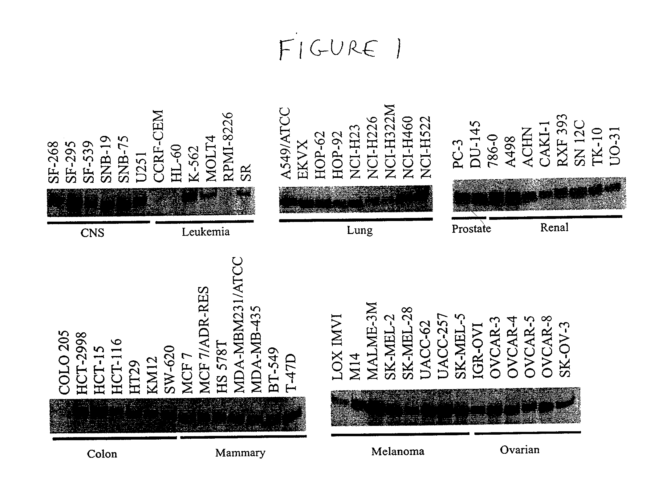
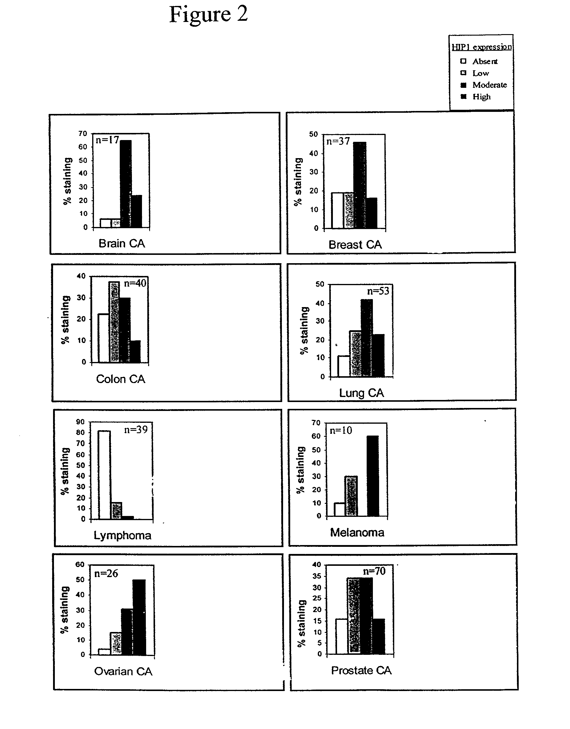
HIP1 Expression in Cancerous and Non-Cancerous Tissues

[0263] A. Monoclonal Antibodies

[0264] The antibodies to HIP1 were generated by isolating a human HIP1 cDNA and creating a bacterial expression vector containing the sequences encoding the carboxyl half of the HIP1 protein. The expressed and purified recombinant protein was use to immunize mice and create monoclonal antibodies by standard methods. The specificity of the antibodies to HIP1 was confirmed by comparing Western blot analyses of murine embryonic fibroblast extracts derived from embryos with HIP1 deleted and their wild type litter mates (data not shown). Three monoclonal antibodies were found to be useful for Western blot analysis of HIP1, as well as for IHC of human tissue. These antibodies were designated HIP1-4B10, HIP1-1A1 and HIP1-1B11. Antibodies 4B10 and 1A1 recognize only human HIP1. Antibody 1B11 recognizes both human and mouse HIP1. The hybridomas producing antibodies HIP1-4B10, 1A1 and HIP1-1B11 were deposited...

example 2

HIP1 Expression in a Mouse Model of Prostate Cancer

[0288] This example describes an investigation of the expression of HIP1 in a transgeric mouse model of prostate cancer, designated the TRAMP mouse (Greenberg et al., PNAS 92:3439 [1995]). The TRAMP mouse was created by use of a transgene where the rat probasin promoter drives expression of SV40 early genes (T and t; Tag). SV40 Tag interacts with p53 and the retinoblasoma gene product Rb abrogating their tumor suppressor gene functions.

[0289] HIP1 was found to be overexpressed in 50% of prostate tumors from TRAMP mice (n=10) compared to the normal prostate (FIG. 6a), suggesting that its expression is an active component of tumorigenesis in these mice. The heterogeneity of HIP1 expression is similar to that seen in human prostate cancers.

example 3

HIP1 ENTH Mutant

[0290] The effect of expression of a HIP1 dominant negative mutant on cellular survival was next investigated. At the molecular level, the dominant negative mutant of HIP1 (HIP1 / deltaE lacking the ENTH domain) promoted cell death but expression of wild type HIP1 (FLHIP1) or vector alone (pcDNA3) did not induce cell death (FIG. 6b). 39% of HIP1 / DE-expressing cells and 50% of caspase 9-expressing cells were apoptotic. FIG. 12 shows the correction of HIP1 deltaE apoptosis by the expression fall length HIP1 (FLPHIP1). FIG. 13 shows the correction of apoptosis by the expression of dominant negative caspase 9 (DN Casp 9) and constitutatively active Akt (Myr-AKT) but not dominant negative caspase 8 (DN I-FLICE) or activated ras (Ras-V12). Expression of the endogenous HIP1, wild type HIP1, HIP1 / DE and Caspase 9 was documented by Western blot analysis as shown in the bottom of FIG. 6b.


PUM

PropertyMeasurementUnit
Compositionaaaaaaaaaa
Cell deathaaaaaaaaaa
Biological propertiesaaaaaaaaaa

Abstract

The present invention relates to compositions and methods for cancer diagnostics and therapeutics, including but not limited to, HIP1 cancer markers. In particular, the present invention provides compositions and methods of using HIP1 in the diagnosis and treatment of epithelial cancers. The present invention thus provides improved compositions and methods for diagnosing and treating some of the most common cancers (e.g., prostate and colon cancers). The present invention additionally provides drugs active against HIP1 and methods for screening for such drugs.

Description

[0001] This application claims priority to Provisional Patent Application Serial No. not yet assigned, filed Nov. 15, 2001 with Express Mail Label EL837033508US.[0003] The present invention relates to compositions and methods for cancer diagnostics, including but not limited to, HIP1 cancer markers. In particular, the present invention provides compositions and methods of using HIP1 in the diagnosis and treatment of epithelial cancers. The present invention further provides methods of screening potential therapeutic compounds for HIP1 inhibitory properties.[0004] Most forms of cancer do not have diagnostic screening tests available. For the cancers that do have screening tests available, the tests are frequently invasive, expensive, and lack strong diagnostic utility.[0005] For example, afflicting one out of nine men over age 65, prostate cancer (PCA) is a leading cause of male cancer-related death, second only to lung cancer (Abate-Shen and Shen, Genes Dev 14:2410 [2000]; Ruijter e...

Claims


Application Information

Patent Timeline
IPC IPC(8): C07K16/30C12P21/08
CPCC07K16/3069C07K16/3046
Inventor ROSS, THEODORAMIZUKAMI, IKUKORAO, DINESH
Owner THE RGT OF THE UNIV OF MICHIGAN
Why Eureka
Try Eureka