
System, method and computer program product for measuring blood properties form a spectral image

a technology of spectral image and system, applied in the field of reflected light analysis, can solve the problems of blood escaping into the tissues, the inability of newborn children, especially premature babies, to effectively process bilirubin,

Inactive Publication Date: 2004-02-05
INTELLIGENT MEDICAL DEVICES
View PDF3 Cites 25 Cited by
  • Summary
  • Abstract
  • Description
  • Claims
  • Application Information

AI Technical Summary

Benefits of technology

[0035] An advantage of the present invention is that it provides a means for the rapid, non-invasive measurement of clinically significant parameters of the CBC+Diff test. It advantageously provides immediate results. As such, it can be used for point-of-care testing and diagnosis.

Problems solved by technology

However, the livers of newborn children, especially premature babies, cannot process bilirubin effectively.
The birth process often results in extensive bruising, resulting in blood escaping into the tissues where it is broken down metabolically.
At still higher levels, deposition begins in deeper tissues, including the brain, and can result in permanent brain damage.
Invasive techniques, such as for conventional CBC+Diff tests and bilirubin analysis, pose particular problems for newborns because their circulatory system is not yet fully developed.
This procedure is traumatic even for an infant in good health.
More importantly, this procedure poses the risk of having to do a blood transfusion because of the low total blood volume of the infant.
In addition to newborns, invasive techniques are also particularly stressful for, and / or difficult to carryout on, children, elderly patients, bum patients, and patients in special care units.
The demarcation between the physical findings of the patient and the laboratory findings are, in general, the result of technical limitations.
However, there are some instances in which pallor may result from other causes, such as constriction of peripheral vessels, or being hidden by skin pigmentation.
Therefore, in vivo visualization of the circulation is difficult because of poor resolution, and generally impractical because of the complexities involved in compensating for multiple scattering and for specular reflection from the surface.
However, the resolution of such images is limited because of the scattering of light, and the computations for the scattering factor are complex.
However, because of its limited range and inaccuracy, reflection spectrophotometry has been used primarily in qualitative rather than quantitative analysis.
However, good Lambertian surfaces are difficult to obtain.
These studies were done to obtain experimental data regarding capillary density, capillary shape, and blood flow velocity, and were limited to gross physical measurements on capillaries.
Because the Winkelman device takes measurements only in capillaries, measurements made by the Winkelman device will not accurately reflect measurements for larger vessels.
This inaccuracy results from the constantly changing relationship of volume of cells to volume of blood in small capillaries resulting from the non-Newtonian viscosity characteristic of blood.
Consequently, the Winkelman device is not capable of measuring the central or true hematocrit, or the total hemoglobin concentration, which depend upon the ratio of the volume of red blood cells to that of the whole blood in a large vessel such as a vein.
However, blood flowing through a micro-capillary will contain approximately 1000 red cells for every white cell, making this an impractical method.
The Winkelman device does not provide any means by which platelets can be visualized and counted.
Further, the Winkelman device does not provide any means by which the capillary plasma can be visualized, or the constituents of the capillary plasma quantified.
The Winkelman device also does not provide a means by which abnormal constituents of blood, such as tumor cells, can be detected.

Method used


Image

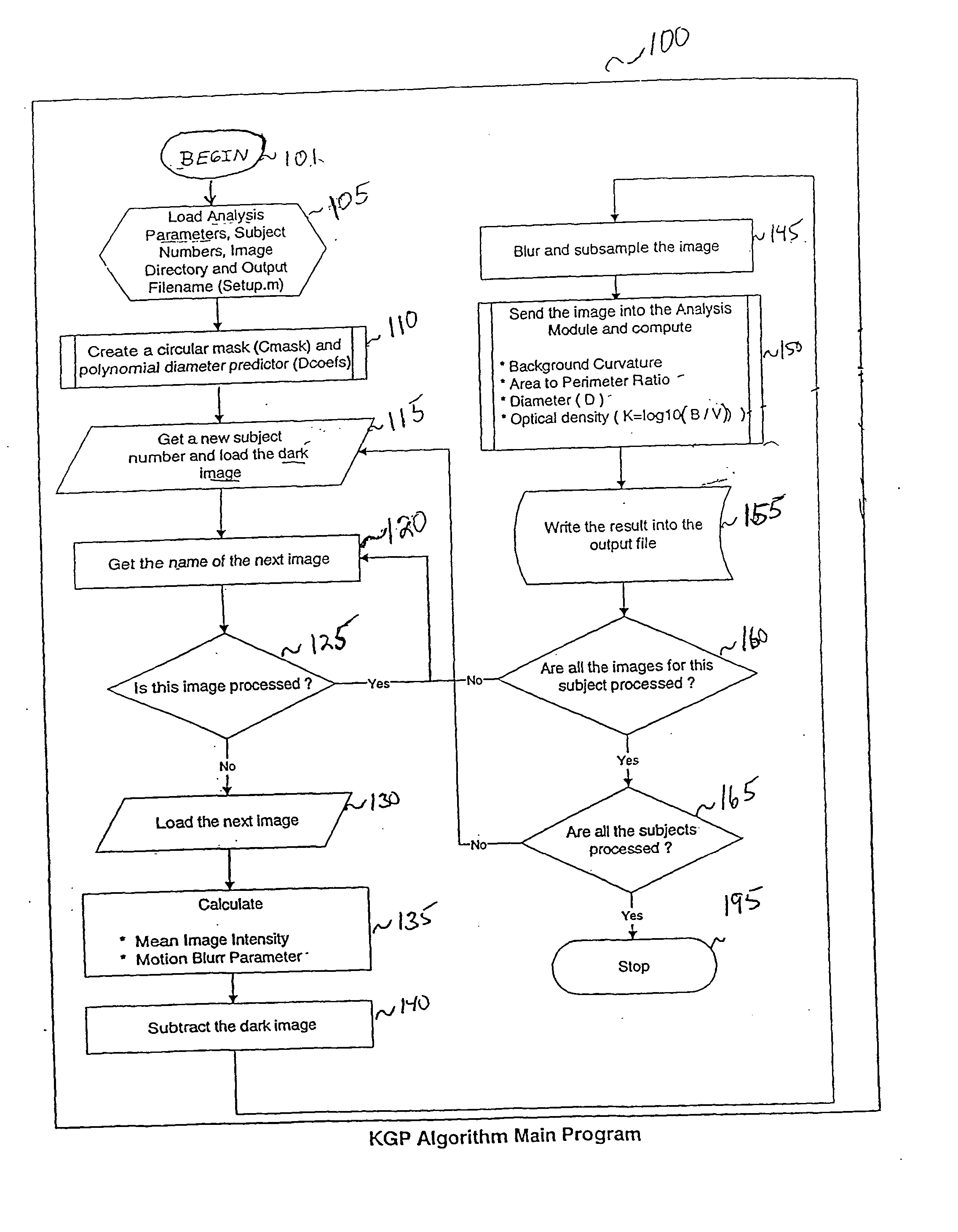
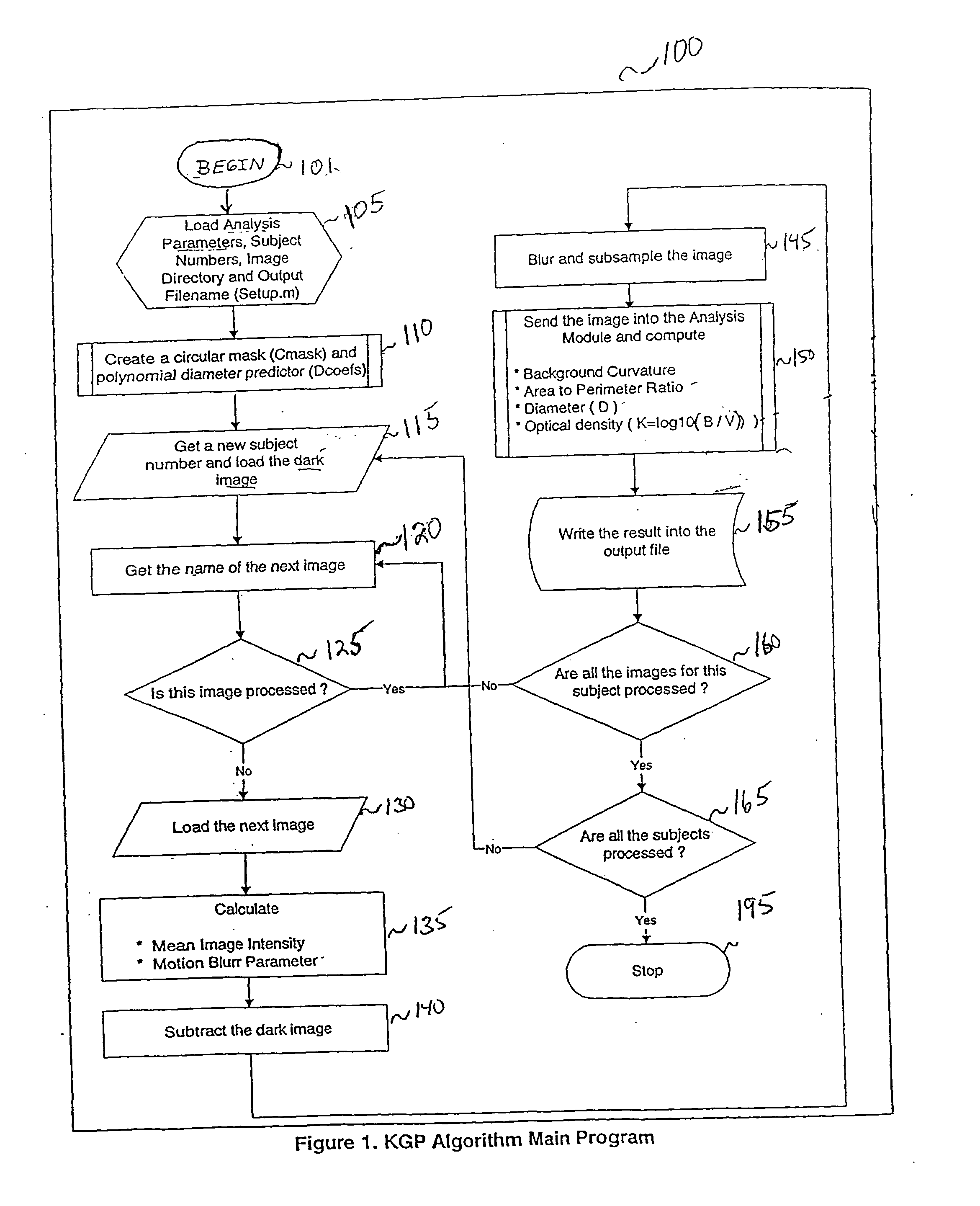
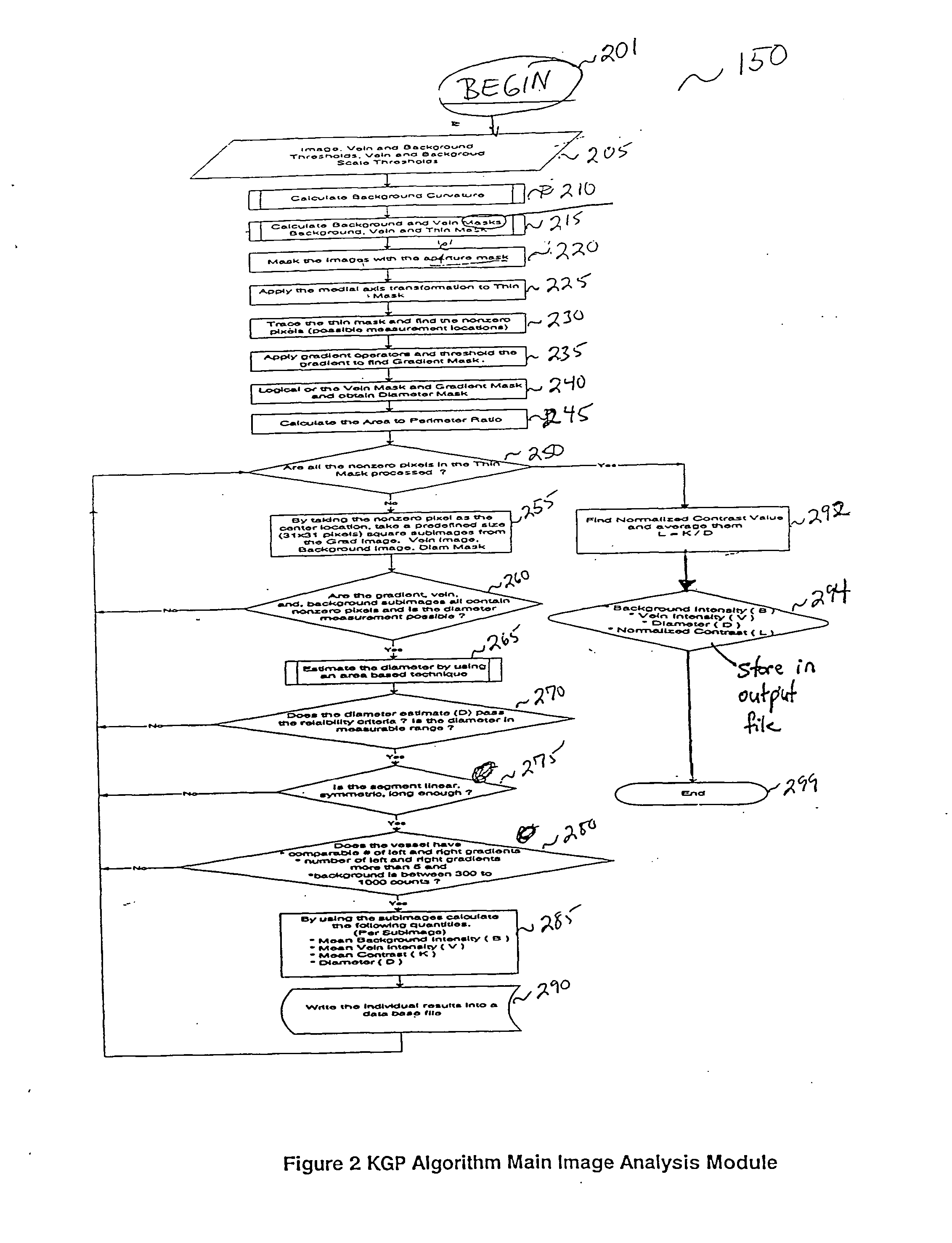
  • System, method and computer program product for measuring blood properties form a spectral image
  • System, method and computer program product for measuring blood properties form a spectral image
  • System, method and computer program product for measuring blood properties form a spectral image

Examples


Embodiment Construction

[0051] I. Overview of the Present Invention

[0052] The present invention is directed to a method and apparatus for analysis, particularly non-invasive, in vivo analysis of a subject's vascular system. The in vivo measurements discussed herein can also be performed in vitro by imaging blood in, for example, a tube or flow cell, as would be apparent to a person skilled in the relevant art(s). The in vivo method is carried out by imaging a portion of the subject's vascular system. For example, the image can be created from a sub-surface region of a subject's tissues or organs. The tissue covering the imaged portion must be traversed by light without multiple scattering to obtain a reflected image. In order to form an image, two criteria must be met. First, there must be image contrast resulting from a difference in the optical properties, such as absorption, index of refraction, or scattering characteristics, between the subject to be imaged and its surroundings or background. Second, t...


PUM


Abstract

A system, method and computer program product is provided for analyzing spectral images of a micrcirculatory system to measure the volume and concentration of a blood vessel. The images are analyzed to identify vessel structure, measure the light absorption and develop a contrast gradient plus (KGP) estimate. The KGP estimate is used to predict blood characteristics, such as hemoglobin concentration and hematocrit. The KGP is estimated in three distinct phases. First, the images are screened to measure a mean image intensity and motion blur. Second, each image is analyzed to identify background curvate, and create vessel, background and diameter masks. To identify background curvate, the images are analyzed to detect shadows caused by larger blood vessels in the background image. The diameter and area of the vessels are also calculated. During the Prediction and Calibration phase, the images are screened to eliminate all images failing thresholds for mean intensity, motion blur, background curvature and area-to-perimeter ratio. Finally, the KGP estimate is determined from the selected images.

Description

[0001] 1. Field of the Invention[0002] The present invention relates generally to reflected light analysis. More particularly, the invention relates to the use of reflected spectral imaging to analyze visualizable components of a fluid flowing in a tubular system. Still more particularly, the invention relates to the use of reflected spectral imaging to analyze the components of blood in a mammalian, especially human, vascular system.[0003] 2. Related Art[0004] Widely accepted medical school doctrine teaches that the complete blood count including the white blood cell differential (CBC+Diff) is one of the best tests to assess a patient's overall health. With it, a physician can detect or diagnose anemia, infection, blood loss, acute and chronic diseases, allergies, and other conditions. CBC+Diff analyses provide comprehensive information on constituents in blood, including the number of red cells, the hematocrit, the hemoglobin concentration, and indices that portray the size, shape...

Claims


Application Information

Patent Timeline
Patent Type & Authority Applications(United States)
IPC IPC(8): A61B5/00
CPCA61B5/0059A61B5/14535A61B5/489A61B5/411A61B5/1455
Inventor COOK, CHRISTOPHER AEMRESOY, MUSTAFA KFARID, HANY
Owner INTELLIGENT MEDICAL DEVICES
Learn More